[English] 日本語
 Yorodumi
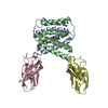
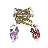
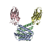
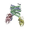
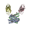
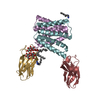
- PDB-7mgx: Structure of EmrE-D3 mutant in complex with monobody L10 and meth... -
+ Open data
- Basic information
| Entry | Database: PDB / ID: 7mgx | ||||||
|---|---|---|---|---|---|---|---|
| Title | Structure of EmrE-D3 mutant in complex with monobody L10 and methyl viologen | ||||||
 Components Components |
| ||||||
 Keywords Keywords | TRANSPORT PROTEIN/IMMUNE SYSTEM / small multidrug resistance transporter / paraquat / TRANSPORT PROTEIN / TRANSPORT PROTEIN-IMMUNE SYSTEM complex | ||||||
| Function / homology |  Function and homology information Function and homology informationEmrE multidrug transporter complex / amino-acid betaine transmembrane transporter activity / glycine betaine transport / choline transmembrane transporter activity / choline transport / xenobiotic detoxification by transmembrane export across the plasma membrane / antiporter activity / response to osmotic stress / xenobiotic transport / xenobiotic transmembrane transporter activity ...EmrE multidrug transporter complex / amino-acid betaine transmembrane transporter activity / glycine betaine transport / choline transmembrane transporter activity / choline transport / xenobiotic detoxification by transmembrane export across the plasma membrane / antiporter activity / response to osmotic stress / xenobiotic transport / xenobiotic transmembrane transporter activity / transmembrane transporter activity / xenobiotic metabolic process / transmembrane transport / cellular response to xenobiotic stimulus / response to xenobiotic stimulus / DNA damage response / identical protein binding / membrane / plasma membrane Similarity search - Function | ||||||
| Biological species |   Homo sapiens (human) Homo sapiens (human) | ||||||
| Method |  X-RAY DIFFRACTION / X-RAY DIFFRACTION /  SYNCHROTRON / SYNCHROTRON /  MOLECULAR REPLACEMENT / Resolution: 3.13 Å MOLECULAR REPLACEMENT / Resolution: 3.13 Å | ||||||
 Authors Authors | Kermani, A.A. / Stockbridge, R.B. | ||||||
| Funding support |  United States, 1items United States, 1items
| ||||||
 Citation Citation |  Journal: Elife / Year: 2022 Journal: Elife / Year: 2022Title: Crystal structures of bacterial small multidrug resistance transporter EmrE in complex with structurally diverse substrates. Authors: Kermani, A.A. / Burata, O.E. / Koff, B.B. / Koide, A. / Koide, S. / Stockbridge, R.B. | ||||||
| History |
|
- Structure visualization
| Structure viewer | Molecule:  Molmil Molmil Jmol/JSmol Jmol/JSmol |
|---|
- Downloads & links
- Download
| PDBx/mmCIF format |  7mgx.cif.gz 7mgx.cif.gz | 154.9 KB | Display |  PDBx/mmCIF format PDBx/mmCIF format |
|---|---|---|---|---|
| PDB format |  pdb7mgx.ent.gz pdb7mgx.ent.gz | 122.4 KB | Display |  PDB format PDB format |
| PDBx/mmJSON format |  7mgx.json.gz 7mgx.json.gz | Tree view |  PDBx/mmJSON format PDBx/mmJSON format | |
| Others |  Other downloads Other downloads |
-Validation report
| Summary document |  7mgx_validation.pdf.gz 7mgx_validation.pdf.gz | 821.8 KB | Display |  wwPDB validaton report wwPDB validaton report |
|---|---|---|---|---|
| Full document |  7mgx_full_validation.pdf.gz 7mgx_full_validation.pdf.gz | 845.2 KB | Display | |
| Data in XML |  7mgx_validation.xml.gz 7mgx_validation.xml.gz | 28.9 KB | Display | |
| Data in CIF |  7mgx_validation.cif.gz 7mgx_validation.cif.gz | 38.4 KB | Display | |
| Arichive directory |  https://data.pdbj.org/pub/pdb/validation_reports/mg/7mgx https://data.pdbj.org/pub/pdb/validation_reports/mg/7mgx ftp://data.pdbj.org/pub/pdb/validation_reports/mg/7mgx ftp://data.pdbj.org/pub/pdb/validation_reports/mg/7mgx | HTTPS FTP |
-Related structure data
| Related structure data |  7mh6SC 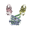 7ssuC 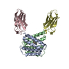 7sv9C 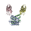 7svxC  7sztC  7t00C S: Starting model for refinement C: citing same article ( |
|---|---|
| Similar structure data |
- Links
Links
- Assembly
| Deposited unit | 
| ||||||||
|---|---|---|---|---|---|---|---|---|---|
| 1 | 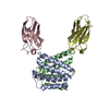
| ||||||||
| 2 | 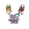
| ||||||||
| Unit cell |
|
- Components
| #1: Protein | Mass: 11907.279 Da / Num. of mol.: 4 / Mutation: E25N, W31I, V34M Source method: isolated from a genetically manipulated source Source: (gene. exp.)   #2: Antibody | Mass: 9931.934 Da / Num. of mol.: 4 Source method: isolated from a genetically manipulated source Source: (gene. exp.)  Homo sapiens (human) / Production host: Homo sapiens (human) / Production host:  #3: Chemical | Has ligand of interest | Y | |
|---|
-Experimental details
-Experiment
| Experiment | Method:  X-RAY DIFFRACTION / Number of used crystals: 1 X-RAY DIFFRACTION / Number of used crystals: 1 |
|---|
- Sample preparation
| Crystal | Density Matthews: 4.47 Å3/Da / Density % sol: 72.51 % |
|---|---|
| Crystal grow | Temperature: 298 K / Method: vapor diffusion, sitting drop / Details: 0.1 M NH4SO4, 0.1 M ADA, pH 6.3, 35% PEG600 |
-Data collection
| Diffraction | Mean temperature: 80 K / Serial crystal experiment: N |
|---|---|
| Diffraction source | Source:  SYNCHROTRON / Site: SYNCHROTRON / Site:  APS APS  / Beamline: 21-ID-D / Wavelength: 0.987 Å / Beamline: 21-ID-D / Wavelength: 0.987 Å |
| Detector | Type: DECTRIS EIGER X 9M / Detector: PIXEL / Date: Feb 11, 2021 |
| Radiation | Monochromator: Si(111) / Protocol: SINGLE WAVELENGTH / Monochromatic (M) / Laue (L): M / Scattering type: x-ray |
| Radiation wavelength | Wavelength: 0.987 Å / Relative weight: 1 |
| Reflection | Resolution: 3.13→70.839 Å / Num. obs: 14289 / % possible obs: 82 % / Redundancy: 3.7 % / CC1/2: 0.939 / Rmerge(I) obs: 0.123 / Rrim(I) all: 0.1 / Net I/σ(I): 7.7 |
| Reflection shell | Resolution: 3.13→3.419 Å / Num. unique obs: 714 / CC1/2: 0.629 / % possible all: 72.3 |
- Processing
| Software |
| ||||||||||||||||||||||||
|---|---|---|---|---|---|---|---|---|---|---|---|---|---|---|---|---|---|---|---|---|---|---|---|---|---|
| Refinement | Method to determine structure:  MOLECULAR REPLACEMENT MOLECULAR REPLACEMENTStarting model: 7MH6 Resolution: 3.13→32.92 Å / SU ML: 0.6 / Cross valid method: THROUGHOUT / σ(F): 1.97 / Phase error: 46.88 / Stereochemistry target values: ML
| ||||||||||||||||||||||||
| Solvent computation | Shrinkage radii: 0.9 Å / VDW probe radii: 1.11 Å / Solvent model: FLAT BULK SOLVENT MODEL | ||||||||||||||||||||||||
| Displacement parameters | Biso max: 155.19 Å2 / Biso mean: 74.0881 Å2 / Biso min: 36.01 Å2 | ||||||||||||||||||||||||
| Refinement step | Cycle: final / Resolution: 3.13→32.92 Å
| ||||||||||||||||||||||||
| LS refinement shell | Resolution: 3.13→3.24 Å / Rfactor Rfree error: 0
|
 Movie
Movie Controller





 PDBj
PDBj


