 Movie
Movie Controller


+ Open data

- Basic information
| Entry | Database: PDB / ID: 7m0h | ||||||
|---|---|---|---|---|---|---|---|
| Title | DHP B in complex with 4-chlorophenol ligand | ||||||
 Components Components | Dehaloperoxidase B | ||||||
 Keywords Keywords | OXIDOREDUCTASE / peroxidase / peroxygenase / complex | ||||||
| Function / homology |  Function and homology information Function and homology informationoxygen carrier activity / peroxidase activity / oxygen binding / heme binding / metal ion binding Similarity search - Function | ||||||
| Biological species |   Amphitrite ornata (invertebrata) Amphitrite ornata (invertebrata) | ||||||
| Method |  X-RAY DIFFRACTION / SYNCHROTRON / MOLECULAR REPLACEMENT / Resolution: 1.91 Å X-RAY DIFFRACTION / SYNCHROTRON / MOLECULAR REPLACEMENT / Resolution: 1.91 Å | ||||||
 Authors Authors | Carey, L.M. / Ghiladi, R.A. | ||||||
| Funding support |  United States, 1items United States, 1items
| ||||||
 Citation Citation | Journal: To Be Published Title: Mechanistic and Structural Studies of 2,4-dihalophenol: Bridging the Functional Gap between Reactivity and Inhibition in Dehaloperoxidase Authors: Carey, L.M. / Ghiladi, R.A. | ||||||
| History |
|
- Structure visualization
| Structure viewer | Molecule: MolmilJmol/JSmol |
|---|
- Downloads & links
-Download
| PDBx/mmCIF format | 7m0h.cif.gz | 74.8 KB | Display | PDBx/mmCIF format |
|---|---|---|---|---|
| PDB format | pdb7m0h.ent.gz | 54.7 KB | Display | PDB format |
| PDBx/mmJSON format | 7m0h.json.gz | Tree view | PDBx/mmJSON format | |
| Others |  Other downloads Other downloads |
-Validation report
| Arichive directory | https://data.pdbj.org/pub/pdb/validation_reports/m0/7m0hftp://data.pdbj.org/pub/pdb/validation_reports/m0/7m0h | HTTPS FTP |
|---|
-Related structure data
| Related structure data |  7lzkC  7lznC  7lzoC  7m0fC  3ixfS C: citing same article ( S: Starting model for refinement |
|---|---|
| Similar structure data |
-Links
 PDBj
PDBj


- Assembly
| Deposited unit | 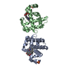
| ||||||||
|---|---|---|---|---|---|---|---|---|---|
| 1 | 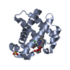
| ||||||||
| 2 | 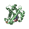
| ||||||||
| Unit cell |
|
-Components
| #1: Protein | Mass: 15414.462 Da / Num. of mol.: 2 Source method: isolated from a genetically manipulated source Source: (gene. exp.) Amphitrite ornata (invertebrata) / Plasmid: pET16bProduction host:  References: UniProt: Q9NAV7 #2: Chemical |   Mass: 616.487 Da / Num. of mol.: 2 Mass: 616.487 Da / Num. of mol.: 2Source method: isolated from a genetically manipulated source Formula: C34H32FeN4O4 #3: Chemical |   Mass: 96.063 Da / Num. of mol.: 2 / Source method: obtained synthetically / Formula: SO4 Mass: 96.063 Da / Num. of mol.: 2 / Source method: obtained synthetically / Formula: SO4#4: Chemical |   Mass: 128.556 Da / Num. of mol.: 2 / Source method: obtained synthetically / Formula: C6H5ClO Mass: 128.556 Da / Num. of mol.: 2 / Source method: obtained synthetically / Formula: C6H5ClO#5: Water | ChemComp-HOH / |  Mass: 18.015 Da / Num. of mol.: 184 / Source method: isolated from a natural source / Formula: H2O Mass: 18.015 Da / Num. of mol.: 184 / Source method: isolated from a natural source / Formula: H2OHas ligand of interest | N | |
|---|
-Experimental details
-Experiment
| Experiment | Method: X-RAY DIFFRACTION / Number of used crystals: 1 |
|---|
- Sample preparation
| Crystal | Density Matthews: 2.12 Å3/Da / Density % sol: 42.03 % |
|---|---|
| Crystal grow | Temperature: 277 K / Method: vapor diffusion, hanging drop / pH: 6.4 Details: PEG4000, ammonium sulfate, sodium cacodylate buffer |
-Data collection
| Diffraction | Mean temperature: 100 K / Serial crystal experiment: N |
|---|---|
| Diffraction source | Source: SYNCHROTRON / Site: APS / Beamline: 22-BM / Wavelength: 1 Å |
| Detector | Type: MARMOSAIC 225 mm CCD / Detector: CCD / Date: Aug 13, 2015 |
| Radiation | Protocol: SINGLE WAVELENGTH / Monochromatic (M) / Laue (L): M / Scattering type: x-ray |
| Radiation wavelength | Wavelength: 1 Å / Relative weight: 1 |
| Reflection | Resolution: 1.91→50 Å / Num. obs: 21635 / % possible obs: 99.6 % / Redundancy: 4.8 % / Rmerge(I) obs: 0.081 / Net I/σ(I): 14.6 |
| Reflection shell | Resolution: 1.92→1.95 Å / Redundancy: 4.7 % / Rmerge(I) obs: 0.565 / Mean I/σ(I) obs: 1.9 / Num. unique obs: 1047 / CC1/2: 0.761 / % possible all: 99.6 |
- Processing
| Software |
| |||||||||||||||||||||||||||||||||||||||||||||||||||||||||||||||||||||||||||||||||||||||||||||||||||||||||
|---|---|---|---|---|---|---|---|---|---|---|---|---|---|---|---|---|---|---|---|---|---|---|---|---|---|---|---|---|---|---|---|---|---|---|---|---|---|---|---|---|---|---|---|---|---|---|---|---|---|---|---|---|---|---|---|---|---|---|---|---|---|---|---|---|---|---|---|---|---|---|---|---|---|---|---|---|---|---|---|---|---|---|---|---|---|---|---|---|---|---|---|---|---|---|---|---|---|---|---|---|---|---|---|---|---|---|
| Refinement | Method to determine structure: MOLECULAR REPLACEMENT Starting model: 3IXF Resolution: 1.91→37.264 Å / SU ML: 0.21 / Cross valid method: FREE R-VALUE / σ(F): 1.35 / Phase error: 22.43 / Stereochemistry target values: ML
| |||||||||||||||||||||||||||||||||||||||||||||||||||||||||||||||||||||||||||||||||||||||||||||||||||||||||
| Solvent computation | Shrinkage radii: 0.9 Å / VDW probe radii: 1.11 Å / Solvent model: FLAT BULK SOLVENT MODEL | |||||||||||||||||||||||||||||||||||||||||||||||||||||||||||||||||||||||||||||||||||||||||||||||||||||||||
| Refinement step | Cycle: LAST / Resolution: 1.91→37.264 Å
| |||||||||||||||||||||||||||||||||||||||||||||||||||||||||||||||||||||||||||||||||||||||||||||||||||||||||
| Refine LS restraints |
| |||||||||||||||||||||||||||||||||||||||||||||||||||||||||||||||||||||||||||||||||||||||||||||||||||||||||
| LS refinement shell |
|
