[English] 日本語
Yorodumi
- PDB-7lz0: Structure of glutamate receptor-like channel GLR3.4 ligand-bindin... -

+
Open data


ID or keywords:

Loading...

-
Basic information

Entry
Database: PDB / ID: 7lz0
TitleStructure of glutamate receptor-like channel GLR3.4 ligand-binding domain in complex with glutamate
ComponentsGlutamate receptor 3.4
KeywordsMEMBRANE PROTEIN / Arabidopsis thaliana / Ion-Channel / glutamate receptor-like channel (GLR) / Ligand binding domain
Function / homology
Function and homology information


cellular response to acetate / chloroplast membrane / glutamate receptor activity / cellular response to cold / plastid / ligand-gated monoatomic ion channel activity / chloroplast / calcium-mediated signaling / cellular response to amino acid stimulus / calcium channel activity ...cellular response to acetate / chloroplast membrane / glutamate receptor activity / cellular response to cold / plastid / ligand-gated monoatomic ion channel activity / chloroplast / calcium-mediated signaling / cellular response to amino acid stimulus / calcium channel activity / response to wounding / cellular response to mechanical stimulus / calcium ion transport / plasma membrane
Similarity search - Function
Ionotropic glutamate receptor, plant / Plant glutamate receptor, periplasmic ligand-binding domain / Bacterial extracellular solute-binding proteins, family 3 / Solute-binding protein family 3/N-terminal domain of MltF / : / Ligand-gated ion channel / Ionotropic glutamate receptor, L-glutamate and glycine-binding domain / Ligated ion channel L-glutamate- and glycine-binding site / Ionotropic glutamate receptor / Eukaryotic homologues of bacterial periplasmic substrate binding proteins. ...Ionotropic glutamate receptor, plant / Plant glutamate receptor, periplasmic ligand-binding domain / Bacterial extracellular solute-binding proteins, family 3 / Solute-binding protein family 3/N-terminal domain of MltF / : / Ligand-gated ion channel / Ionotropic glutamate receptor, L-glutamate and glycine-binding domain / Ligated ion channel L-glutamate- and glycine-binding site / Ionotropic glutamate receptor / Eukaryotic homologues of bacterial periplasmic substrate binding proteins. / Receptor, ligand binding region / Receptor family ligand binding region / Periplasmic binding protein-like I
Similarity search - Domain/homology
GLUTAMIC ACID / Glutamate receptor 3.4
Similarity search - Component
Biological speciesArabidopsis thaliana (thale cress)
MethodX-RAY DIFFRACTION / SYNCHROTRON / MOLECULAR REPLACEMENT / molecular replacement / Resolution: 2.29 Å
AuthorsGangwar, S.P. / Green, M.N. / Sobolevsky, A.I.
Funding support United States, 4items
OrganizationGrant numberCountry
National Institutes of Health/National Cancer Institute (NIH/NCI)R01 CA206573 United States
National Institutes of Health/National Institute of Neurological Disorders and Stroke (NIH/NINDS)R01 NS083660 United States
National Institutes of Health/National Institute of Neurological Disorders and Stroke (NIH/NINDS)R01 NS107253 United States
National Science Foundation (NSF, United States)1818086 United States
Citation
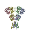
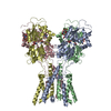
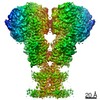
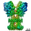
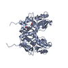
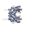
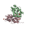
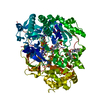
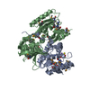
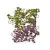
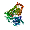
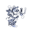
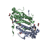
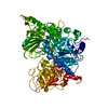
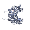
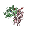
Journal: Mol Cell / Year: 2021
Title: Structure of the Arabidopsis thaliana glutamate receptor-like channel GLR3.4.
Authors: Marriah N Green / Shanti Pal Gangwar / Erwan Michard / Alexander A Simon / Maria Teresa Portes / Juan Barbosa-Caro / Michael M Wudick / Michael A Lizzio / Oleg Klykov / Maria V Yelshanskaya ...Authors: Marriah N Green / Shanti Pal Gangwar / Erwan Michard / Alexander A Simon / Maria Teresa Portes / Juan Barbosa-Caro / Michael M Wudick / Michael A Lizzio / Oleg Klykov / Maria V Yelshanskaya / José A Feijó / Alexander I Sobolevsky /
Abstract: Glutamate receptor-like channels (GLRs) play vital roles in various physiological processes in plants, such as wound response, stomatal aperture control, seed germination, root development, innate ...Glutamate receptor-like channels (GLRs) play vital roles in various physiological processes in plants, such as wound response, stomatal aperture control, seed germination, root development, innate immune response, pollen tube growth, and morphogenesis. Despite the importance of GLRs, knowledge about their molecular organization is limited. Here we use X-ray crystallography and single-particle cryo-EM to solve structures of the Arabidopsis thaliana GLR3.4. Our structures reveal the tetrameric assembly of GLR3.4 subunits into a three-layer domain architecture, reminiscent of animal ionotropic glutamate receptors (iGluRs). However, the non-swapped arrangement between layers of GLR3.4 domains, binding of glutathione through S-glutathionylation of cysteine C205 inside the amino-terminal domain clamshell, unique symmetry, inter-domain interfaces, and ligand specificity distinguish GLR3.4 from representatives of the iGluR family and suggest distinct features of the GLR gating mechanism. Our work elaborates on the principles of GLR architecture and symmetry and provides a molecular template for deciphering GLR-dependent signaling mechanisms in plants.
History
DepositionMar 8, 2021Deposition site: RCSB / Processing site: RCSB
Revision 1.0Jul 28, 2021Provider: repository / Type: Initial release
Revision 1.1Aug 18, 2021Group: Advisory / Database references
Category: citation / database_2 / pdbx_unobs_or_zero_occ_atoms
Item: _citation.journal_volume / _citation.page_first ..._citation.journal_volume / _citation.page_first / _database_2.pdbx_DOI / _database_2.pdbx_database_accession
Revision 1.2Oct 18, 2023Group: Data collection / Refinement description
Category: chem_comp_atom / chem_comp_bond ...chem_comp_atom / chem_comp_bond / pdbx_initial_refinement_model / struct_ncs_dom_lim
Item: _struct_ncs_dom_lim.beg_auth_comp_id / _struct_ncs_dom_lim.beg_label_asym_id ..._struct_ncs_dom_lim.beg_auth_comp_id / _struct_ncs_dom_lim.beg_label_asym_id / _struct_ncs_dom_lim.beg_label_comp_id / _struct_ncs_dom_lim.beg_label_seq_id / _struct_ncs_dom_lim.end_auth_comp_id / _struct_ncs_dom_lim.end_label_asym_id / _struct_ncs_dom_lim.end_label_comp_id / _struct_ncs_dom_lim.end_label_seq_id

-
Structure visualization

Structure viewerMolecule:
MolmilJmol/JSmol

Downloads & links

-
Assembly

Deposited unit
A: Glutamate receptor 3.4
B: Glutamate receptor 3.4
C: Glutamate receptor 3.4
hetero molecules


Theoretical massNumber of molelcules
Total (without water)90,49612
Polymers89,6033
Non-polymers8939
Water1,76598
1
A: Glutamate receptor 3.4
hetero molecules

A: Glutamate receptor 3.4
hetero molecules


Theoretical massNumber of molelcules
Total (without water)60,0304
Polymers59,7352
Non-polymers2942
Water362
TypeNameSymmetry operationNumber
identity operation1_555x,y,z1
crystal symmetry operation12_554x,x-y,-z-1/61
2
B: Glutamate receptor 3.4
C: Glutamate receptor 3.4
hetero molecules


Theoretical massNumber of molelcules
Total (without water)60,48110
Polymers59,7352
Non-polymers7458
Water362
TypeNameSymmetry operationNumber
identity operation1_555x,y,z1
Unit cell
Length a, b, c (Å)74.337, 74.337, 513.560
Angle α, β, γ (deg.)90.000, 90.000, 120.000
Int Tables number179
Space group name H-MP6522
Noncrystallographic symmetry (NCS)NCS domain:
IDEns-IDDetails
11A
21B
12A
22C
13B
23C

NCS domain segments:

Component-ID: 0 / Refine code: 0

Dom-IDEns-IDBeg auth comp-IDBeg label comp-IDEnd auth comp-IDEnd label comp-IDAuth asym-IDLabel asym-IDAuth seq-IDLabel seq-ID
11SERSERCYSCYSAA6 - 25122 - 267
21SERSERCYSCYSBB6 - 25122 - 267
12GLYGLYSERSERAA1 - 25217 - 268
22GLYGLYSERSERCC1 - 25217 - 268
13SERSERCYSCYSBB6 - 25122 - 267
23SERSERCYSCYSCC6 - 25122 - 267

NCS ensembles :
ID
1
2
3

-
Components

-
Protein , 1 types, 3 molecules ABC

#1: Protein Glutamate receptor 3.4 / AtGLR3.4 / Glutamate receptor-like protein 3.4 / Ligand-gated ion channel 3.4


Mass: 29867.660 Da / Num. of mol.: 3 / Fragment: UNP residues 492-601,709-842
Source method: isolated from a genetically manipulated source
Source: (gene. exp.) Arabidopsis thaliana (thale cress) / Gene: GLR3.4, GLR4, GLUR3, At1g05200, YUP8H12.19 / Plasmid: pET22b / Production host: Escherichia coli BL21(DE3) (bacteria) / Strain (production host): Origami (DE3) / References: UniProt: Q8GXJ4

-
Non-polymers , 5 types, 107 molecules

#2: Chemical ChemComp-GLU / GLUTAMIC ACID / Glutamic acid


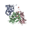
Type: L-peptide linking / Mass: 147.129 Da / Num. of mol.: 3 / Source method: obtained synthetically / Formula: C5H9NO4 / Feature type: SUBJECT OF INVESTIGATION
#3: Chemical ChemComp-CL / CHLORIDE ION / Chloride


Mass: 35.453 Da / Num. of mol.: 2 / Source method: obtained synthetically / Formula: Cl
#4: Chemical ChemComp-SO4 / SULFATE ION / Sulfate


Mass: 96.063 Da / Num. of mol.: 3 / Source method: obtained synthetically / Formula: SO4
#5: Chemical ChemComp-GOL / GLYCEROL / GLYCERIN / PROPANE-1,2,3-TRIOL / Glycerol


Mass: 92.094 Da / Num. of mol.: 1 / Source method: obtained synthetically / Formula: C3H8O3
#6: Water ChemComp-HOH / water / Water


Mass: 18.015 Da / Num. of mol.: 98 / Source method: isolated from a natural source / Formula: H2O

-
Details

Has ligand of interestY

-
Experimental details

-
Experiment

ExperimentMethod: X-RAY DIFFRACTION / Number of used crystals: 1

-
Sample preparation

CrystalDensity Matthews: 2.49 Å3/Da / Density % sol: 50.69 % / Description: small octagonal crystals
Crystal growTemperature: 277 K / Method: vapor diffusion / pH: 4.6 / Details: 2 M ammonium sulfate, 0.1 M sodium acetate, pH 4.6 / Temp details: Cold room

-
Data collection

DiffractionMean temperature: 100 K / Ambient temp details: Liquid Nitrogen / Serial crystal experiment: N
Diffraction sourceSource: SYNCHROTRON / Site: APS / Beamline: 24-ID-C / Wavelength: 0.9791 Å
DetectorType: DECTRIS PILATUS 6M-F / Detector: PIXEL / Date: Nov 6, 2019
RadiationMonochromator: cryo-cooled double crystal Si(111) / Protocol: SINGLE WAVELENGTH / Monochromatic (M) / Laue (L): M / Scattering type: x-ray
Radiation wavelengthWavelength: 0.9791 Å / Relative weight: 1
ReflectionResolution: 2.29→64.46 Å / Num. obs: 39519 / % possible obs: 99.7 % / Redundancy: 9.3 % / CC1/2: 0.998 / Rmerge(I) obs: 0.123 / Rpim(I) all: 0.042 / Rrim(I) all: 0.13 / Net I/σ(I): 12.9
Reflection shell

Diffraction-ID: 1

Resolution (Å)Redundancy (%)Rmerge(I) obsNum. measured allNum. unique obsCC1/2Rpim(I) allRrim(I) allNet I/σ(I) obs% possible all
2.29-2.379.20.8093363236570.8050.2750.8562.897
8.88-64.3870.02862178850.9990.0110.0331.299.6

-
Phasing

PhasingMethod: molecular replacement

-
Processing

Software
NameVersionClassificationNB
XDSdata reduction
Aimless0.7.4data scaling
PHASERphasing
REFMAC5.8.0267refinement
PDB_EXTRACT3.27data extraction
RefinementMethod to determine structure: MOLECULAR REPLACEMENT
Starting model: PDB entry 6VEA
Resolution: 2.29→64.46 Å / Cor.coef. Fo:Fc: 0.944 / Cor.coef. Fo:Fc free: 0.928 / SU B: 7.435 / SU ML: 0.175 / Cross valid method: THROUGHOUT / σ(F): 0 / ESU R: 0.329 / ESU R Free: 0.227 / Stereochemistry target values: MAXIMUM LIKELIHOOD
Details: HYDROGENS HAVE BEEN ADDED IN THE RIDING POSITIONS U VALUES: REFINED INDIVIDUALLY
RfactorNum. reflection% reflectionSelection details
Rfree0.2426 1962 5 %RANDOM
Rwork0.2095 ---
obs0.2112 37406 99.62 %-
Solvent computationIon probe radii: 0.8 Å / Shrinkage radii: 0.8 Å / VDW probe radii: 1.2 Å / Solvent model: MASK
Displacement parametersBiso max: 124.04 Å2 / Biso mean: 36.822 Å2 / Biso min: 21.31 Å2
Baniso -1Baniso -2Baniso -3
1--0.61 Å2-0.31 Å20 Å2
2---0.61 Å2-0 Å2
3---1.99 Å2
Refinement stepCycle: final / Resolution: 2.29→64.46 Å
ProteinNucleic acidLigandSolventTotal
Num. atoms5877 0 53 98 6028
Biso mean--42.51 30.68 -
Num. residues----751
Refine LS restraints
Refine-IDTypeDev idealDev ideal targetNumber
X-RAY DIFFRACTIONr_bond_refined_d0.0040.0136042
X-RAY DIFFRACTIONr_bond_other_d0.0010.0175712
X-RAY DIFFRACTIONr_angle_refined_deg1.2511.6498221
X-RAY DIFFRACTIONr_angle_other_deg1.1451.57913142
X-RAY DIFFRACTIONr_dihedral_angle_1_deg6.6875743
X-RAY DIFFRACTIONr_dihedral_angle_2_deg31.37822.546326
X-RAY DIFFRACTIONr_dihedral_angle_3_deg14.41315977
X-RAY DIFFRACTIONr_dihedral_angle_4_deg12.6571542
X-RAY DIFFRACTIONr_chiral_restr0.0520.2788
X-RAY DIFFRACTIONr_gen_planes_refined0.0040.026863
X-RAY DIFFRACTIONr_gen_planes_other0.0010.021365
Refine LS restraints NCS

Refine-ID: X-RAY DIFFRACTION / Type: interatomic distance / Weight position: 0.05

Ens-IDDom-IDAuth asym-IDNumberRms dev position (Å)
11A75080.12
12B75080.12
21A76460.11
22C76460.11
31B76670.11
32C76670.11
LS refinement shellResolution: 2.293→2.353 Å / Rfactor Rfree error: 0 / Total num. of bins used: 20
RfactorNum. reflection% reflection
Rfree0.297 135 -
Rwork0.276 2589 -
all-2724 -
obs--95.95 %

+
About Yorodumi

-
News

-
Feb 9, 2022. New format data for meta-information of EMDB entries

New format data for meta-information of EMDB entries

  • Version 3 of the EMDB header file is now the official format.
  • The previous official version 1.9 will be removed from the archive.

Related info.:EMDB header

External links:wwPDB to switch to version 3 of the EMDB data model

-
Aug 12, 2020. Covid-19 info

Covid-19 info

URL: https://pdbj.org/emnavi/covid19.php

New page: Covid-19 featured information page in EM Navigator.

Related info.:Covid-19 info / Mar 5, 2020. Novel coronavirus structure data

+
Mar 5, 2020. Novel coronavirus structure data

Novel coronavirus structure data

Related info.:Yorodumi Speices / Aug 12, 2020. Covid-19 info

External links:COVID-19 featured content - PDBj / Molecule of the Month (242):Coronavirus Proteases

+
Jan 31, 2019. EMDB accession codes are about to change! (news from PDBe EMDB page)

EMDB accession codes are about to change! (news from PDBe EMDB page)

  • The allocation of 4 digits for EMDB accession codes will soon come to an end. Whilst these codes will remain in use, new EMDB accession codes will include an additional digit and will expand incrementally as the available range of codes is exhausted. The current 4-digit format prefixed with “EMD-” (i.e. EMD-XXXX) will advance to a 5-digit format (i.e. EMD-XXXXX), and so on. It is currently estimated that the 4-digit codes will be depleted around Spring 2019, at which point the 5-digit format will come into force.
  • The EM Navigator/Yorodumi systems omit the EMD- prefix.

Related info.:Q: What is EMD? / ID/Accession-code notation in Yorodumi/EM Navigator

External links:EMDB Accession Codes are Changing Soon! / Contact to PDBj

+
Jul 12, 2017. Major update of PDB

Major update of PDB

  • wwPDB released updated PDB data conforming to the new PDBx/mmCIF dictionary.
  • This is a major update changing the version number from 4 to 5, and with Remediation, in which all the entries are updated.
  • In this update, many items about electron microscopy experimental information are reorganized (e.g. em_software).
  • Now, EM Navigator and Yorodumi are based on the updated data.

External links:wwPDB Remediation / Enriched Model Files Conforming to OneDep Data Standards Now Available in the PDB FTP Archive

-
Yorodumi

Thousand views of thousand structures

  • Yorodumi is a browser for structure data from EMDB, PDB, SASBDB, etc.
  • This page is also the successor to EM Navigator detail page, and also detail information page/front-end page for Omokage search.
  • The word "yorodu" (or yorozu) is an old Japanese word meaning "ten thousand". "mi" (miru) is to see.

Related info.:EMDB / PDB / SASBDB / Comparison of 3 databanks / Yorodumi Search / Aug 31, 2016. New EM Navigator & Yorodumi / Yorodumi Papers / Jmol/JSmol / Function and homology information / Changes in new EM Navigator and Yorodumi

Read more