 Movie
Movie Controller

[English] 日本語
 Yorodumi
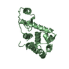
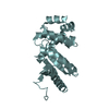
- PDB-7luh: Burkholderia pseudomallei Disulfide bond forming protein A (DsbA)... -
+ Open data

- Basic information
| Entry | Database: PDB / ID: 7luh | ||||||||||||
|---|---|---|---|---|---|---|---|---|---|---|---|---|---|
| Title | Burkholderia pseudomallei Disulfide bond forming protein A (DsbA) liganded with fragment bromophenoxy propanamide | ||||||||||||
 Components Components | Thiol:disulfide interchange protein | ||||||||||||
 Keywords Keywords | OXIDOREDUCTASE / BpsDsbA / Fragment / cryptic-pocket | ||||||||||||
| Function / homology |  Function and homology information Function and homology information | ||||||||||||
| Biological species |  Burkholderia pseudomallei (bacteria) Burkholderia pseudomallei (bacteria) | ||||||||||||
| Method |  X-RAY DIFFRACTION / SYNCHROTRON / MOLECULAR REPLACEMENT / Resolution: 1.84 Å X-RAY DIFFRACTION / SYNCHROTRON / MOLECULAR REPLACEMENT / Resolution: 1.84 Å | ||||||||||||
 Authors Authors | Petit, G.A. / Martin, J.L. / McMahon, R.M. | ||||||||||||
| Funding support |  Australia, 3items Australia, 3items
| ||||||||||||
 Citation Citation | Journal: Acta Crystallogr D Struct Biol / Year: 2022 Title: Identification and characterization of two drug-like fragments that bind to the same cryptic binding pocket of Burkholderia pseudomallei DsbA. Authors: Petit, G.A. / Mohanty, B. / McMahon, R.M. / Nebl, S. / Hilko, D.H. / Wilde, K.L. / Scanlon, M.J. / Martin, J.L. / Halili, M.A. | ||||||||||||
| History |
|
- Structure visualization
| Structure viewer | Molecule: MolmilJmol/JSmol |
|---|
- Downloads & links
-Download
| PDBx/mmCIF format | 7luh.cif.gz | 156.1 KB | Display | PDBx/mmCIF format |
|---|---|---|---|---|
| PDB format | pdb7luh.ent.gz | 101.6 KB | Display | PDB format |
| PDBx/mmJSON format | 7luh.json.gz | Tree view | PDBx/mmJSON format | |
| Others |  Other downloads Other downloads |
-Validation report
| Summary document | 7luh_validation.pdf.gz | 685.6 KB | Display | wwPDB validaton report |
|---|---|---|---|---|
| Full document | 7luh_full_validation.pdf.gz | 685.9 KB | Display | |
| Data in XML | 7luh_validation.xml.gz | 11.5 KB | Display | |
| Data in CIF | 7luh_validation.cif.gz | 16.7 KB | Display | |
| Arichive directory | https://data.pdbj.org/pub/pdb/validation_reports/lu/7luhftp://data.pdbj.org/pub/pdb/validation_reports/lu/7luh | HTTPS FTP |
-Related structure data
| Related structure data |  7lujC  4k2dS S: Starting model for refinement C: citing same article ( |
|---|---|
| Similar structure data |


-Links
 PDBj
PDBj

- Assembly
| Deposited unit | 
| ||||||||||||
|---|---|---|---|---|---|---|---|---|---|---|---|---|---|
| 1 |
| ||||||||||||
| Unit cell |
|
-Components
| #1: Protein | Mass: 22022.115 Da / Num. of mol.: 1 Source method: isolated from a genetically manipulated source Details: The 3 first amino acids are left over from TEV cleavage scar Source: (gene. exp.) Burkholderia pseudomallei (strain K96243) (bacteria)Strain: K96243 / Gene: dsbA, BPSL0381 / Production host: |
|---|---|
| #2: Chemical | ChemComp-YCS / (  Mass: 244.085 Da / Num. of mol.: 1 / Source method: obtained synthetically / Formula: C9H10BrNO2 / Feature type: SUBJECT OF INVESTIGATION Mass: 244.085 Da / Num. of mol.: 1 / Source method: obtained synthetically / Formula: C9H10BrNO2 / Feature type: SUBJECT OF INVESTIGATION |
| #3: Water | ChemComp-HOH /  Mass: 18.015 Da / Num. of mol.: 206 / Source method: isolated from a natural source / Formula: H2O Mass: 18.015 Da / Num. of mol.: 206 / Source method: isolated from a natural source / Formula: H2O |
| Has ligand of interest | Y |
| Has protein modification | Y |
-Experimental details
-Experiment
| Experiment | Method: X-RAY DIFFRACTION / Number of used crystals: 1 |
|---|
- Sample preparation
| Crystal | Density Matthews: 3.03 Å3/Da / Density % sol: 59.35 % / Description: Long needles, transparent |
|---|---|
| Crystal grow | Temperature: 293.15 K / Method: vapor diffusion, sitting drop / pH: 7.5 / Details: 60% Tacsimate Cryo protected in 20% Ethylen Glycol |
-Data collection
| Diffraction | Mean temperature: 100 K / Serial crystal experiment: N |
|---|---|
| Diffraction source | Source: SYNCHROTRON / Site: Australian Synchrotron / Beamline: MX2 / Wavelength: 0.9537 Å |
| Detector | Type: DECTRIS EIGER X 9M / Detector: PIXEL / Date: Nov 22, 2019 |
| Radiation | Protocol: SINGLE WAVELENGTH / Monochromatic (M) / Laue (L): M / Scattering type: x-ray |
| Radiation wavelength | Wavelength: 0.9537 Å / Relative weight: 1 |
| Reflection | Resolution: 1.84→46.62 Å / Num. obs: 23090 / % possible obs: 98.75 % / Redundancy: 6.7 % / Biso Wilson estimate: 31.21 Å2 / CC1/2: 0.999 / CC star: 1 / Rmerge(I) obs: 0.071 / Rpim(I) all: 0.03 / Rrim(I) all: 0.077 / Net I/σ(I): 16 |
| Reflection shell | Resolution: 1.84→1.91 Å / Redundancy: 2 % / Rmerge(I) obs: 1.27 / Mean I/σ(I) obs: 1.51 / Num. unique obs: 2249 / CC1/2: 0.633 / CC star: 0.88 / Rpim(I) all: 0.512 / Rrim(I) all: 1.37 / % possible all: 98.38 |
- Processing
| Software |
| |||||||||||||||||||||||||||||||||||||||||||||||||||||||||||||||||||||||||||||||||||||||||||||||||||||||||
|---|---|---|---|---|---|---|---|---|---|---|---|---|---|---|---|---|---|---|---|---|---|---|---|---|---|---|---|---|---|---|---|---|---|---|---|---|---|---|---|---|---|---|---|---|---|---|---|---|---|---|---|---|---|---|---|---|---|---|---|---|---|---|---|---|---|---|---|---|---|---|---|---|---|---|---|---|---|---|---|---|---|---|---|---|---|---|---|---|---|---|---|---|---|---|---|---|---|---|---|---|---|---|---|---|---|---|
| Refinement | Method to determine structure: MOLECULAR REPLACEMENT Starting model: 4K2D Resolution: 1.84→46.61 Å / SU ML: 0.2374 / Cross valid method: FREE R-VALUE / σ(F): 1.35 / Phase error: 20.0311 Stereochemistry target values: GeoStd + Monomer Library + CDL v1.2 Details: Cycle of automated refinement with Phenix refine, using geometry and ADP weight optimisation. TLS (3 groups: residues 7-70, 71-137 and 138-197). Occupancy of the ligand was set to 0.5 and ...Details: Cycle of automated refinement with Phenix refine, using geometry and ADP weight optimisation. TLS (3 groups: residues 7-70, 71-137 and 138-197). Occupancy of the ligand was set to 0.5 and then refined using occupancy refinement. Manual refinement using coot V 0.9.5
| |||||||||||||||||||||||||||||||||||||||||||||||||||||||||||||||||||||||||||||||||||||||||||||||||||||||||
| Solvent computation | Shrinkage radii: 0.9 Å / VDW probe radii: 1.11 Å / Solvent model: FLAT BULK SOLVENT MODEL | |||||||||||||||||||||||||||||||||||||||||||||||||||||||||||||||||||||||||||||||||||||||||||||||||||||||||
| Displacement parameters | Biso mean: 37.15 Å2 | |||||||||||||||||||||||||||||||||||||||||||||||||||||||||||||||||||||||||||||||||||||||||||||||||||||||||
| Refinement step | Cycle: LAST / Resolution: 1.84→46.61 Å
| |||||||||||||||||||||||||||||||||||||||||||||||||||||||||||||||||||||||||||||||||||||||||||||||||||||||||
| Refine LS restraints |
| |||||||||||||||||||||||||||||||||||||||||||||||||||||||||||||||||||||||||||||||||||||||||||||||||||||||||
| LS refinement shell |
| |||||||||||||||||||||||||||||||||||||||||||||||||||||||||||||||||||||||||||||||||||||||||||||||||||||||||
| Refinement TLS params. | Method: refined / Refine-ID: X-RAY DIFFRACTION
| |||||||||||||||||||||||||||||||||||||||||||||||||||||||||||||||||||||||||||||||||||||||||||||||||||||||||
| Refinement TLS group | Refine-ID: X-RAY DIFFRACTION / Auth asym-ID: A / Label asym-ID: A
|
