 Movie
Movie Controller


+ Open data

- Basic information
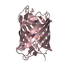
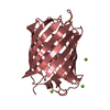
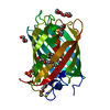
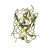
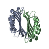
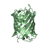
| Entry | Database: PDB / ID: 7lug | |||||||||
|---|---|---|---|---|---|---|---|---|---|---|
| Title | Crystal structure of the pnRFP B30Y mutant | |||||||||
 Components Components | Red Fluorescent pnRFP B30Y mutant | |||||||||
 Keywords Keywords | FLUORESCENT PROTEIN / Red fluorescent protein / Peroxynitrite / Biosensor / Boronophenylalanine. | |||||||||
| Function / homology | Green Fluorescent Protein / Green fluorescent protein / Beta Barrel / Mainly Beta / PHOSPHATE ION Function and homology information Function and homology information | |||||||||
| Biological species |  Discosoma sp. (sea anemone) Discosoma sp. (sea anemone) | |||||||||
| Method |  X-RAY DIFFRACTION / SYNCHROTRON / MOLECULAR REPLACEMENT / Resolution: 1.95 Å X-RAY DIFFRACTION / SYNCHROTRON / MOLECULAR REPLACEMENT / Resolution: 1.95 Å | |||||||||
 Authors Authors | Huang, M. / Ng, H.L. / Pang, Y. / Zhang, S. / Fan, Y. / Yeh, H. / Xiong, Y. / Li, X. / Ai, H. | |||||||||
 Citation Citation | Journal: To Be Published Title: Development, Characterization, and Structural Analysis of a Genetically Encoded Red Fluorescent Peroxynitrite Biosensor Authors: Pang, Y. / Huang, M. / Zhang, S. / Fan, Y. / Yeh, H. / Xiong, Y. / Li, X. / Ng, H.L. / Ai, H. | |||||||||
| History |
|
- Structure visualization
| Structure viewer | Molecule: MolmilJmol/JSmol |
|---|
- Downloads & links
-Download
| PDBx/mmCIF format | 7lug.cif.gz | 61.5 KB | Display | PDBx/mmCIF format |
|---|---|---|---|---|
| PDB format | pdb7lug.ent.gz | 42.9 KB | Display | PDB format |
| PDBx/mmJSON format | 7lug.json.gz | Tree view | PDBx/mmJSON format | |
| Others |  Other downloads Other downloads |
-Validation report
| Arichive directory | https://data.pdbj.org/pub/pdb/validation_reports/lu/7lugftp://data.pdbj.org/pub/pdb/validation_reports/lu/7lug | HTTPS FTP |
|---|
-Related structure data
| Related structure data | 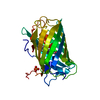 7lqoSC S: Starting model for refinement C: citing same article ( |
|---|---|
| Similar structure data |


-Links
 PDBj
PDBj- Assembly
| Deposited unit | 
| ||||||||
|---|---|---|---|---|---|---|---|---|---|
| 1 |
| ||||||||
| Unit cell |
|
-Components
| #1: Protein | Mass: 29741.689 Da / Num. of mol.: 1 Source method: isolated from a genetically manipulated source Source: (gene. exp.) Discosoma sp. (sea anemone) / Production host:  Escherichia phage EcSzw-2 (virus) Escherichia phage EcSzw-2 (virus) |
|---|---|
| #2: Chemical | ChemComp-PO4 /   Mass: 94.971 Da / Num. of mol.: 1 / Source method: obtained synthetically / Formula: PO4 Mass: 94.971 Da / Num. of mol.: 1 / Source method: obtained synthetically / Formula: PO4 |
| #3: Water | ChemComp-HOH /  Mass: 18.015 Da / Num. of mol.: 65 / Source method: isolated from a natural source / Formula: H2O Mass: 18.015 Da / Num. of mol.: 65 / Source method: isolated from a natural source / Formula: H2O |
| Has ligand of interest | Y |
| Has protein modification | Y |
-Experimental details
-Experiment
| Experiment | Method: X-RAY DIFFRACTION / Number of used crystals: 1 |
|---|
- Sample preparation
| Crystal | Density Matthews: 2.02 Å3/Da / Density % sol: 38.97 % |
|---|---|
| Crystal grow | Temperature: 286 K / Method: vapor diffusion, sitting drop / Details: Sodium phosphate, citric acid, PEG400 |
-Data collection
| Diffraction | Mean temperature: 100 K / Serial crystal experiment: N |
|---|---|
| Diffraction source | Source: SYNCHROTRON / Site: APS  / Beamline: 23-ID-D / Wavelength: 1.03321 Å / Beamline: 23-ID-D / Wavelength: 1.03321 Å |
| Detector | Type: DECTRIS PILATUS3 6M / Detector: PIXEL / Date: Aug 20, 2018 |
| Radiation | Protocol: MAD / Monochromatic (M) / Laue (L): M / Scattering type: x-ray |
| Radiation wavelength | Wavelength: 1.03321 Å / Relative weight: 1 |
| Reflection | Resolution: 1.95→41.85 Å / Num. obs: 17539 / % possible obs: 99.3 % / Redundancy: 3.56 % / CC1/2: 0.998 / Net I/σ(I): 10.8 |
| Reflection shell | Resolution: 1.95→2 Å / Num. unique obs: 1245 / CC1/2: 0.814 |
- Processing
| Software |
| ||||||||||||||||||||||||||||||||||||||||||||||||||||||||||||
|---|---|---|---|---|---|---|---|---|---|---|---|---|---|---|---|---|---|---|---|---|---|---|---|---|---|---|---|---|---|---|---|---|---|---|---|---|---|---|---|---|---|---|---|---|---|---|---|---|---|---|---|---|---|---|---|---|---|---|---|---|---|
| Refinement | Method to determine structure: MOLECULAR REPLACEMENT Starting model: 7LQO Resolution: 1.95→41.85 Å / Cor.coef. Fo:Fc: 0.957 / Cor.coef. Fo:Fc free: 0.95 / SU B: 3.451 / SU ML: 0.1 / Cross valid method: THROUGHOUT / σ(F): 0 / ESU R: 0.172 / ESU R Free: 0.142 / Stereochemistry target values: MAXIMUM LIKELIHOOD Details: HYDROGENS HAVE BEEN ADDED IN THE RIDING POSITIONS U VALUES : REFINED INDIVIDUALLY
| ||||||||||||||||||||||||||||||||||||||||||||||||||||||||||||
| Solvent computation | Ion probe radii: 0.8 Å / Shrinkage radii: 0.8 Å / VDW probe radii: 1.2 Å / Solvent model: MASK | ||||||||||||||||||||||||||||||||||||||||||||||||||||||||||||
| Displacement parameters | Biso max: 66.16 Å2 / Biso mean: 32.588 Å2 / Biso min: 22.66 Å2
| ||||||||||||||||||||||||||||||||||||||||||||||||||||||||||||
| Refinement step | Cycle: final / Resolution: 1.95→41.85 Å
| ||||||||||||||||||||||||||||||||||||||||||||||||||||||||||||
| Refine LS restraints |
| ||||||||||||||||||||||||||||||||||||||||||||||||||||||||||||
| LS refinement shell | Resolution: 1.95→2 Å / Rfactor Rfree error: 0 / Total num. of bins used: 20
|
