 Movie
Movie Controller

[English] 日本語
 Yorodumi
- PDB-7lsd: Crystal structure of near-infrared fluorescent protein miRFP718nano -
+ Open data

- Basic information
| Entry | Database: PDB / ID: 7lsd | ||||||
|---|---|---|---|---|---|---|---|
| Title | Crystal structure of near-infrared fluorescent protein miRFP718nano | ||||||
 Components Components | miRFP718nano | ||||||
 Keywords Keywords | FLUORESCENT PROTEIN / near-infrared fluorescent protein / miRFP / miRFPnano / phytochrome / BphP / cyanobacterichrome / CBCR | ||||||
| Function / homology | GAF domain / Beta-Lactamase / 2-Layer Sandwich / Alpha Beta / BILIVERDINE IX ALPHA Function and homology information Function and homology information | ||||||
| Biological species |  | ||||||
| Method |  X-RAY DIFFRACTION / SYNCHROTRON / MOLECULAR REPLACEMENT / Resolution: 1.7 Å X-RAY DIFFRACTION / SYNCHROTRON / MOLECULAR REPLACEMENT / Resolution: 1.7 Å | ||||||
 Authors Authors | Pletnev, S. | ||||||
 Citation Citation | Journal: To Be Published Title: Rational design of a small near-infrared fluorescent protein from CBCR suitable for deep-tissue SWIR imaging. Authors: Oliinyk, O.S. / Pletnev, S. / Ma, C. / Baloban, M. / Toboada, C. / Sheng, H. / Yao, J. / Verkhusha, V.V. | ||||||
| History |
|
- Structure visualization
| Structure viewer | Molecule: MolmilJmol/JSmol |
|---|
- Downloads & links
-Download
| PDBx/mmCIF format | 7lsd.cif.gz | 156.9 KB | Display | PDBx/mmCIF format |
|---|---|---|---|---|
| PDB format | pdb7lsd.ent.gz | 123.7 KB | Display | PDB format |
| PDBx/mmJSON format | 7lsd.json.gz | Tree view | PDBx/mmJSON format | |
| Others |  Other downloads Other downloads |
-Validation report
| Arichive directory | https://data.pdbj.org/pub/pdb/validation_reports/ls/7lsdftp://data.pdbj.org/pub/pdb/validation_reports/ls/7lsd | HTTPS FTP |
|---|
-Related structure data
| Related structure data | 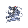 6mghS S: Starting model for refinement |
|---|---|
| Similar structure data |


-Links
 PDBj
PDBj- Assembly
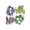
| Deposited unit | 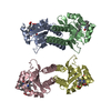
| ||||||||
|---|---|---|---|---|---|---|---|---|---|
| 1 | 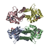
| ||||||||
| Unit cell |
| ||||||||
| Components on special symmetry positions |
|
-Components
| #1: Protein | Mass: 18828.457 Da / Num. of mol.: 4 Source method: isolated from a genetically manipulated source Source: (gene. exp.) #2: Chemical | ChemComp-BLA /   Mass: 582.646 Da / Num. of mol.: 4 / Source method: obtained synthetically / Formula: C33H34N4O6 / Feature type: SUBJECT OF INVESTIGATION Mass: 582.646 Da / Num. of mol.: 4 / Source method: obtained synthetically / Formula: C33H34N4O6 / Feature type: SUBJECT OF INVESTIGATION#3: Water | ChemComp-HOH / |  Mass: 18.015 Da / Num. of mol.: 758 / Source method: isolated from a natural source / Formula: H2O Mass: 18.015 Da / Num. of mol.: 758 / Source method: isolated from a natural source / Formula: H2OHas ligand of interest | Y | Has protein modification | Y | |
|---|
-Experimental details
-Experiment
| Experiment | Method: X-RAY DIFFRACTION / Number of used crystals: 1 |
|---|
- Sample preparation
| Crystal | Density Matthews: 2.72 Å3/Da / Density % sol: 54.73 % |
|---|---|
| Crystal grow | Temperature: 293 K / Method: vapor diffusion, hanging drop / pH: 3.5 Details: 12.6% PEG6000, 0.1 M lithium sulfate, 0.07 M citric acid, pH 3.5, 2.1% D-sorbitol |
-Data collection
| Diffraction | Mean temperature: 100 K / Serial crystal experiment: N | |||||||||||||||||||||||||||||||||||||||||||||||||||||||||||||||||||||||||||||||||||||||||||||||||||
|---|---|---|---|---|---|---|---|---|---|---|---|---|---|---|---|---|---|---|---|---|---|---|---|---|---|---|---|---|---|---|---|---|---|---|---|---|---|---|---|---|---|---|---|---|---|---|---|---|---|---|---|---|---|---|---|---|---|---|---|---|---|---|---|---|---|---|---|---|---|---|---|---|---|---|---|---|---|---|---|---|---|---|---|---|---|---|---|---|---|---|---|---|---|---|---|---|---|---|---|---|
| Diffraction source | Source: SYNCHROTRON / Site: APS  / Beamline: 22-BM / Wavelength: 1 Å / Beamline: 22-BM / Wavelength: 1 Å | |||||||||||||||||||||||||||||||||||||||||||||||||||||||||||||||||||||||||||||||||||||||||||||||||||
| Detector | Type: MARMOSAIC 300 mm CCD / Detector: CCD / Date: Feb 15, 2020 | |||||||||||||||||||||||||||||||||||||||||||||||||||||||||||||||||||||||||||||||||||||||||||||||||||
| Radiation | Monochromator: double crystal Si(111) / Protocol: SINGLE WAVELENGTH / Monochromatic (M) / Laue (L): M / Scattering type: x-ray | |||||||||||||||||||||||||||||||||||||||||||||||||||||||||||||||||||||||||||||||||||||||||||||||||||
| Radiation wavelength | Wavelength: 1 Å / Relative weight: 1 | |||||||||||||||||||||||||||||||||||||||||||||||||||||||||||||||||||||||||||||||||||||||||||||||||||
| Reflection | Resolution: 1.7→30 Å / Num. obs: 80892 / % possible obs: 96.6 % / Redundancy: 3.5 % / Rmerge(I) obs: 0.047 / Rpim(I) all: 0.029 / Rrim(I) all: 0.055 / Χ2: 0.616 / Net I/σ(I): 10.3 / Num. measured all: 285750 | |||||||||||||||||||||||||||||||||||||||||||||||||||||||||||||||||||||||||||||||||||||||||||||||||||
| Reflection shell | Diffraction-ID: 1
|
- Processing
| Software |
| ||||||||||||||||||||||||||||||||||||||||||||||||||||||||||||
|---|---|---|---|---|---|---|---|---|---|---|---|---|---|---|---|---|---|---|---|---|---|---|---|---|---|---|---|---|---|---|---|---|---|---|---|---|---|---|---|---|---|---|---|---|---|---|---|---|---|---|---|---|---|---|---|---|---|---|---|---|---|
| Refinement | Method to determine structure: MOLECULAR REPLACEMENT Starting model: PDB entry 6MGH Resolution: 1.7→29.84 Å / Cor.coef. Fo:Fc: 0.976 / Cor.coef. Fo:Fc free: 0.964 / SU B: 2.358 / SU ML: 0.074 / SU R Cruickshank DPI: 0.0893 / Cross valid method: THROUGHOUT / σ(F): 0 / ESU R: 0.089 / ESU R Free: 0.093 / Stereochemistry target values: MAXIMUM LIKELIHOOD Details: HYDROGENS HAVE BEEN ADDED IN THE RIDING POSITIONS U VALUES : REFINED INDIVIDUALLY
| ||||||||||||||||||||||||||||||||||||||||||||||||||||||||||||
| Solvent computation | Ion probe radii: 0.8 Å / Shrinkage radii: 0.8 Å / VDW probe radii: 1.2 Å / Solvent model: MASK | ||||||||||||||||||||||||||||||||||||||||||||||||||||||||||||
| Displacement parameters | Biso max: 100.38 Å2 / Biso mean: 26.701 Å2 / Biso min: 14.07 Å2
| ||||||||||||||||||||||||||||||||||||||||||||||||||||||||||||
| Refinement step | Cycle: final / Resolution: 1.7→29.84 Å
| ||||||||||||||||||||||||||||||||||||||||||||||||||||||||||||
| Refine LS restraints |
| ||||||||||||||||||||||||||||||||||||||||||||||||||||||||||||
| LS refinement shell | Resolution: 1.7→1.741 Å / Rfactor Rfree error: 0
|
