 Movie
Movie Controller

[English] 日本語
 Yorodumi
- PDB-7lq7: Crystal structure of SARS-CoV-2 receptor binding domain in comple... -
+ Open data

- Basic information
| Entry | Database: PDB / ID: 7lq7 | ||||||
|---|---|---|---|---|---|---|---|
| Title | Crystal structure of SARS-CoV-2 receptor binding domain in complex with antibodies CV503 and COVA1-16 | ||||||
 Components Components |
| ||||||
 Keywords Keywords | IMMUNE SYSTEM / antibody / coronavirus / Fab / spike / COVID-19 / RBD / receptor binding domain | ||||||
| Function / homology |  Function and homology information Function and homology informationMaturation of spike protein / viral translation / Translation of Structural Proteins / Virion Assembly and Release / host cell surface / host extracellular space / suppression by virus of host tetherin activity / Induction of Cell-Cell Fusion / structural constituent of virion / entry receptor-mediated virion attachment to host cell ...Maturation of spike protein / viral translation / Translation of Structural Proteins / Virion Assembly and Release / host cell surface / host extracellular space / suppression by virus of host tetherin activity / Induction of Cell-Cell Fusion / structural constituent of virion / entry receptor-mediated virion attachment to host cell / host cell endoplasmic reticulum-Golgi intermediate compartment membrane / receptor-mediated endocytosis of virus by host cell / membrane fusion / Attachment and Entry / positive regulation of viral entry into host cell / receptor-mediated virion attachment to host cell / receptor ligand activity / host cell surface receptor binding / fusion of virus membrane with host plasma membrane / fusion of virus membrane with host endosome membrane / viral envelope / virion attachment to host cell / SARS-CoV-2 activates/modulates innate and adaptive immune responses / host cell plasma membrane / virion membrane / identical protein binding / membrane / plasma membrane Similarity search - Function | ||||||
| Biological species |   Severe acute respiratory syndrome coronavirus 2 Severe acute respiratory syndrome coronavirus 2 Homo sapiens (human) Homo sapiens (human) | ||||||
| Method |  X-RAY DIFFRACTION / SYNCHROTRON / MOLECULAR REPLACEMENT / Resolution: 3.4 Å X-RAY DIFFRACTION / SYNCHROTRON / MOLECULAR REPLACEMENT / Resolution: 3.4 Å | ||||||
 Authors Authors | Yuan, M. / Zhu, X. / Wilson, I.A. | ||||||
| Funding support |  United States, 1items United States, 1items
| ||||||
 Citation Citation | Journal: Sci Transl Med / Year: 2021 Title: Bispecific antibodies targeting distinct regions of the spike protein potently neutralize SARS-CoV-2 variants of concern. Authors: Cho, H. / Gonzales-Wartz, K.K. / Huang, D. / Yuan, M. / Peterson, M. / Liang, J. / Beutler, N. / Torres, J.L. / Cong, Y. / Postnikova, E. / Bangaru, S. / Talana, C.A. / Shi, W. / Yang, E.S. ...Authors: Cho, H. / Gonzales-Wartz, K.K. / Huang, D. / Yuan, M. / Peterson, M. / Liang, J. / Beutler, N. / Torres, J.L. / Cong, Y. / Postnikova, E. / Bangaru, S. / Talana, C.A. / Shi, W. / Yang, E.S. / Zhang, Y. / Leung, K. / Wang, L. / Peng, L. / Skinner, J. / Li, S. / Wu, N.C. / Liu, H. / Dacon, C. / Moyer, T. / Cohen, M. / Zhao, M. / Lee, F.E. / Weinberg, R.S. / Douagi, I. / Gross, R. / Schmaljohn, C. / Pegu, A. / Mascola, J.R. / Holbrook, M. / Nemazee, D. / Rogers, T.F. / Ward, A.B. / Wilson, I.A. / Crompton, P.D. / Tan, J. | ||||||
| History |
|
- Structure visualization
| Structure viewer | Molecule: MolmilJmol/JSmol |
|---|
- Downloads & links
-Download
| PDBx/mmCIF format | 7lq7.cif.gz | 614 KB | Display | PDBx/mmCIF format |
|---|---|---|---|---|
| PDB format | pdb7lq7.ent.gz | 498.9 KB | Display | PDB format |
| PDBx/mmJSON format | 7lq7.json.gz | Tree view | PDBx/mmJSON format | |
| Others |  Other downloads Other downloads |
-Validation report
| Summary document | 7lq7_validation.pdf.gz | 545.8 KB | Display | wwPDB validaton report |
|---|---|---|---|---|
| Full document | 7lq7_full_validation.pdf.gz | 575 KB | Display | |
| Data in XML | 7lq7_validation.xml.gz | 104.6 KB | Display | |
| Data in CIF | 7lq7_validation.cif.gz | 142.9 KB | Display | |
| Arichive directory | https://data.pdbj.org/pub/pdb/validation_reports/lq/7lq7ftp://data.pdbj.org/pub/pdb/validation_reports/lq/7lq7 | HTTPS FTP |
-Related structure data
| Related structure data | 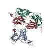 7jmwS S: Starting model for refinement |
|---|---|
| Similar structure data |


-Links
 PDBj
PDBj


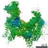
- Assembly
| Deposited unit | 
| |||||||||||||||||||||||||||||||||||||||||||||||||||||||||||||||||||||||||||||||||||||||||||||||||||||||||||||||||||||||||||||||||||||||||||||||||||||||||||||||||||||||||||||||||||||||||||||||||||||||||||||||||||||||||||||||||||||||||||||||||||||||||||||||||||||||||||||||||||||||||||||
|---|---|---|---|---|---|---|---|---|---|---|---|---|---|---|---|---|---|---|---|---|---|---|---|---|---|---|---|---|---|---|---|---|---|---|---|---|---|---|---|---|---|---|---|---|---|---|---|---|---|---|---|---|---|---|---|---|---|---|---|---|---|---|---|---|---|---|---|---|---|---|---|---|---|---|---|---|---|---|---|---|---|---|---|---|---|---|---|---|---|---|---|---|---|---|---|---|---|---|---|---|---|---|---|---|---|---|---|---|---|---|---|---|---|---|---|---|---|---|---|---|---|---|---|---|---|---|---|---|---|---|---|---|---|---|---|---|---|---|---|---|---|---|---|---|---|---|---|---|---|---|---|---|---|---|---|---|---|---|---|---|---|---|---|---|---|---|---|---|---|---|---|---|---|---|---|---|---|---|---|---|---|---|---|---|---|---|---|---|---|---|---|---|---|---|---|---|---|---|---|---|---|---|---|---|---|---|---|---|---|---|---|---|---|---|---|---|---|---|---|---|---|---|---|---|---|---|---|---|---|---|---|---|---|---|---|---|---|---|---|---|---|---|---|---|---|---|---|---|---|---|---|---|---|---|---|---|---|---|---|---|---|---|---|---|---|---|---|---|---|---|---|---|---|---|---|---|---|---|---|---|---|---|---|---|---|---|
| 1 |
| |||||||||||||||||||||||||||||||||||||||||||||||||||||||||||||||||||||||||||||||||||||||||||||||||||||||||||||||||||||||||||||||||||||||||||||||||||||||||||||||||||||||||||||||||||||||||||||||||||||||||||||||||||||||||||||||||||||||||||||||||||||||||||||||||||||||||||||||||||||||||||||
| Unit cell |
| |||||||||||||||||||||||||||||||||||||||||||||||||||||||||||||||||||||||||||||||||||||||||||||||||||||||||||||||||||||||||||||||||||||||||||||||||||||||||||||||||||||||||||||||||||||||||||||||||||||||||||||||||||||||||||||||||||||||||||||||||||||||||||||||||||||||||||||||||||||||||||||
| Noncrystallographic symmetry (NCS) | NCS domain:
NCS domain segments: Component-ID: _ / Refine code: _
|