 Movie
Movie Controller

[English] 日本語
 Yorodumi
- PDB-7loj: T4 lysozyme mutant L99A in complex with 4-(3-phenylpropyl)aniline -
+ Open data

- Basic information
| Entry | Database: PDB / ID: 7loj | ||||||
|---|---|---|---|---|---|---|---|
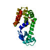
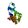
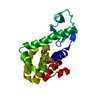
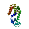
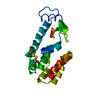
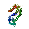
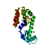
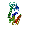
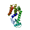
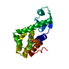
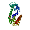
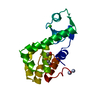
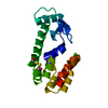
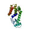
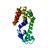
| Title | T4 lysozyme mutant L99A in complex with 4-(3-phenylpropyl)aniline | ||||||
 Components Components | Lysozyme | ||||||
 Keywords Keywords | PROTEIN BINDING / HYDROLASE / mutant / lysozyme / small molecule / L99A / complex | ||||||
| Function / homology |  Function and homology information Function and homology informationviral release from host cell by cytolysis / peptidoglycan catabolic process / cell wall macromolecule catabolic process / lysozyme / lysozyme activity / host cell cytoplasm / defense response to bacterium Similarity search - Function | ||||||
| Biological species |  Enterobacteria phage T4 (virus) Enterobacteria phage T4 (virus) | ||||||
| Method |  X-RAY DIFFRACTION / SYNCHROTRON / MOLECULAR REPLACEMENT / molecular replacement / Resolution: 1.5 Å X-RAY DIFFRACTION / SYNCHROTRON / MOLECULAR REPLACEMENT / molecular replacement / Resolution: 1.5 Å | ||||||
 Authors Authors | Kamenik, A.S. / Singh, I. / Lak, P. / Balius, T.E. / Liedl, K.R. / Shoichet, B.K. | ||||||
| Funding support |  United States, 1items United States, 1items
| ||||||
 Citation Citation | Journal: Proc.Natl.Acad.Sci.USA / Year: 2021 Title: Energy penalties enhance flexible receptor docking in a model cavity. Authors: Kamenik, A.S. / Singh, I. / Lak, P. / Balius, T.E. / Liedl, K.R. / Shoichet, B.K. | ||||||
| History |
|
- Structure visualization
| Structure viewer | Molecule: MolmilJmol/JSmol |
|---|
- Downloads & links
-Download
| PDBx/mmCIF format | 7loj.cif.gz | 52.6 KB | Display | PDBx/mmCIF format |
|---|---|---|---|---|
| PDB format | pdb7loj.ent.gz | 34.2 KB | Display | PDB format |
| PDBx/mmJSON format | 7loj.json.gz | Tree view | PDBx/mmJSON format | |
| Others |  Other downloads Other downloads |
-Validation report
| Arichive directory | https://data.pdbj.org/pub/pdb/validation_reports/lo/7lojftp://data.pdbj.org/pub/pdb/validation_reports/lo/7loj | HTTPS FTP |
|---|
-Related structure data
| Related structure data |  7loaC  7lobC  7locC  7lodC  7loeC 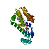 7lofC  7logC  7lx6C  7lx7C  7lx8C 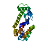 7lx9C  7lxaC 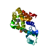 4w57S S: Starting model for refinement C: citing same article ( |
|---|---|
| Similar structure data |


-Links
 PDBj
PDBj


- Assembly
| Deposited unit | 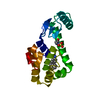
| ||||||||
|---|---|---|---|---|---|---|---|---|---|
| 1 |
| ||||||||
| Unit cell |
|
-Components
| #1: Protein | Mass: 19691.541 Da / Num. of mol.: 1 / Mutation: L99A Source method: isolated from a genetically manipulated source Source: (gene. exp.) Enterobacteria phage T4 (virus) / Gene: e, T4Tp126Production host:  References: UniProt: D9IEF7, lysozyme |
|---|---|
| #2: Chemical | ChemComp-EPE /   Mass: 238.305 Da / Num. of mol.: 1 / Source method: obtained synthetically / Formula: C8H18N2O4S / Comment: pH buffer*YM Mass: 238.305 Da / Num. of mol.: 1 / Source method: obtained synthetically / Formula: C8H18N2O4S / Comment: pH buffer*YM |
| #3: Chemical | ChemComp-Y8M /   Mass: 211.302 Da / Num. of mol.: 1 / Source method: obtained synthetically / Formula: C15H17N / Feature type: SUBJECT OF INVESTIGATION Mass: 211.302 Da / Num. of mol.: 1 / Source method: obtained synthetically / Formula: C15H17N / Feature type: SUBJECT OF INVESTIGATION |
| #4: Water | ChemComp-HOH /  Mass: 18.015 Da / Num. of mol.: 139 / Source method: isolated from a natural source / Formula: H2O Mass: 18.015 Da / Num. of mol.: 139 / Source method: isolated from a natural source / Formula: H2O |
| Has ligand of interest | Y |
-Experimental details
-Experiment
| Experiment | Method: X-RAY DIFFRACTION / Number of used crystals: 1 |
|---|
- Sample preparation
| Crystal | Density Matthews: 2.56 Å3/Da / Density % sol: 52.02 % |
|---|---|
| Crystal grow | Temperature: 294 K / Method: vapor diffusion, hanging drop / pH: 8 Details: Isopropanol, PEG 4000, Tris-Cl pH 8.0, Beta-mercaptoethanol, 2-hyrdoxyethyl disulfide |
-Data collection
| Diffraction | Mean temperature: 100 K / Serial crystal experiment: N | ||||||||||||||||||||||||||||||
|---|---|---|---|---|---|---|---|---|---|---|---|---|---|---|---|---|---|---|---|---|---|---|---|---|---|---|---|---|---|---|---|
| Diffraction source | Source: SYNCHROTRON / Site: ALS / Beamline: 8.3.1 / Wavelength: 1.11583 Å | ||||||||||||||||||||||||||||||
| Detector | Type: DECTRIS PILATUS3 S 6M / Detector: PIXEL / Date: Nov 11, 2018 | ||||||||||||||||||||||||||||||
| Radiation | Monochromator: M / Protocol: SINGLE WAVELENGTH / Monochromatic (M) / Laue (L): M / Scattering type: x-ray | ||||||||||||||||||||||||||||||
| Radiation wavelength | Wavelength: 1.11583 Å / Relative weight: 1 | ||||||||||||||||||||||||||||||
| Reflection | Resolution: 1.5→52.31 Å / Num. obs: 32996 / % possible obs: 99.7 % / Redundancy: 8.5 % / Biso Wilson estimate: 13.73 Å2 / CC1/2: 0.998 / Rmerge(I) obs: 0.036 / Rpim(I) all: 0.013 / Rrim(I) all: 0.038 / Net I/σ(I): 30.8 / Num. measured all: 281442 / Scaling rejects: 252 | ||||||||||||||||||||||||||||||
| Reflection shell | Diffraction-ID: 1
|
-Phasing
| Phasing | Method: molecular replacement |
|---|
- Processing
| Software |
| ||||||||||||||||||||||||||||||||||||||||||||||||||||||||||||||||||||||||
|---|---|---|---|---|---|---|---|---|---|---|---|---|---|---|---|---|---|---|---|---|---|---|---|---|---|---|---|---|---|---|---|---|---|---|---|---|---|---|---|---|---|---|---|---|---|---|---|---|---|---|---|---|---|---|---|---|---|---|---|---|---|---|---|---|---|---|---|---|---|---|---|---|---|
| Refinement | Method to determine structure: MOLECULAR REPLACEMENT Starting model: 4W57 Resolution: 1.5→52.31 Å / SU ML: 0.12 / Cross valid method: THROUGHOUT / σ(F): 1.44 / Phase error: 19.18 / Stereochemistry target values: ML
| ||||||||||||||||||||||||||||||||||||||||||||||||||||||||||||||||||||||||
| Solvent computation | Shrinkage radii: 0.9 Å / VDW probe radii: 1.11 Å / Solvent model: FLAT BULK SOLVENT MODEL | ||||||||||||||||||||||||||||||||||||||||||||||||||||||||||||||||||||||||
| Displacement parameters | Biso max: 51.63 Å2 / Biso mean: 16.0541 Å2 / Biso min: 8.48 Å2 | ||||||||||||||||||||||||||||||||||||||||||||||||||||||||||||||||||||||||
| Refinement step | Cycle: final / Resolution: 1.5→52.31 Å
| ||||||||||||||||||||||||||||||||||||||||||||||||||||||||||||||||||||||||
| Refine LS restraints |
| ||||||||||||||||||||||||||||||||||||||||||||||||||||||||||||||||||||||||
| LS refinement shell | Refine-ID: X-RAY DIFFRACTION / Rfactor Rfree error: 0
|
