 Movie
Movie Controller

[English] 日本語
 Yorodumi
- PDB-7lnq: Structure of the avibactam-CDD-1 3 minute complex in imidazole and MPD -
+ Open data

- Basic information
| Entry | Database: PDB / ID: 7lnq | ||||||
|---|---|---|---|---|---|---|---|
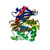
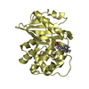
| Title | Structure of the avibactam-CDD-1 3 minute complex in imidazole and MPD | ||||||
 Components Components | Beta-lactamase | ||||||
 Keywords Keywords | HYDROLASE/INHIBITOR / beta-lactamase / antibiotic resistance / HYDROLASE / HYDROLASE-INHIBITOR complex | ||||||
| Function / homology |  Function and homology information Function and homology informationpenicillin binding / antibiotic catabolic process / beta-lactamase activity / beta-lactamase / response to antibiotic / membrane Similarity search - Function | ||||||
| Biological species |  Clostridioides difficile (bacteria) Clostridioides difficile (bacteria) | ||||||
| Method |  X-RAY DIFFRACTION / SYNCHROTRON / FOURIER SYNTHESIS / Resolution: 1.73 Å X-RAY DIFFRACTION / SYNCHROTRON / FOURIER SYNTHESIS / Resolution: 1.73 Å | ||||||
 Authors Authors | Smith, C.A. / Vakulenko, S.B. / Stewart, N.K. | ||||||
 Citation Citation | Journal: Acs Infect Dis. / Year: 2021 Title: In Crystallo Time-Resolved Interaction of the Clostridioides difficile CDD-1 enzyme with Avibactam Provides New Insights into the Catalytic Mechanism of Class D beta-lactamases. Authors: Stewart, N.K. / Toth, M. / Stasyuk, A. / Vakulenko, S.B. / Smith, C.A. | ||||||
| History |
|
- Structure visualization
| Structure viewer | Molecule: MolmilJmol/JSmol |
|---|
- Downloads & links
-Download
| PDBx/mmCIF format | 7lnq.cif.gz | 74 KB | Display | PDBx/mmCIF format |
|---|---|---|---|---|
| PDB format | pdb7lnq.ent.gz | 52.4 KB | Display | PDB format |
| PDBx/mmJSON format | 7lnq.json.gz | Tree view | PDBx/mmJSON format | |
| Others |  Other downloads Other downloads |
-Validation report
| Arichive directory | https://data.pdbj.org/pub/pdb/validation_reports/ln/7lnqftp://data.pdbj.org/pub/pdb/validation_reports/ln/7lnq | HTTPS FTP |
|---|
-Related structure data
| Related structure data |  7lnoC 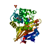 7lnrC  7me9C  7meaC  7mebC  7mecC  7medC  7meeC  7mefC  7megC  7mehC  6edmS S: Starting model for refinement C: citing same article ( |
|---|---|
| Similar structure data |


-Links
 PDBj
PDBj
- Assembly
| Deposited unit | 
| |||||||||
|---|---|---|---|---|---|---|---|---|---|---|
| 1 |
| |||||||||
| Unit cell |
| |||||||||
| Components on special symmetry positions |
|
-Components
-Protein , 1 types, 1 molecules A
| #1: Protein | Mass: 36043.055 Da / Num. of mol.: 1 / Mutation: K238A, K244A Source method: isolated from a genetically manipulated source Source: (gene. exp.) Clostridioides difficile (bacteria)Gene: blaR1_4, blaR1_1, E5F39_11445, SAMEA2239407_03320, SAMEA3374989_01677 Production host: |
|---|
-Non-polymers , 5 types, 187 molecules 


| #2: Chemical | ChemComp-NXL / ( Mass: 267.260 Da / Num. of mol.: 1 / Source method: obtained synthetically / Formula: C7H13N3O6S / Feature type: SUBJECT OF INVESTIGATION / Comment: antibiotic*YM Mass: 267.260 Da / Num. of mol.: 1 / Source method: obtained synthetically / Formula: C7H13N3O6S / Feature type: SUBJECT OF INVESTIGATION / Comment: antibiotic*YM | ||||
|---|---|---|---|---|---|
| #3: Chemical | ChemComp-CO2 /  Mass: 44.010 Da / Num. of mol.: 1 / Source method: obtained synthetically / Formula: CO2 Mass: 44.010 Da / Num. of mol.: 1 / Source method: obtained synthetically / Formula: CO2 | ||||
| #4: Chemical | ChemComp-SO4 /  Mass: 96.063 Da / Num. of mol.: 4 / Source method: obtained synthetically / Formula: SO4 Mass: 96.063 Da / Num. of mol.: 4 / Source method: obtained synthetically / Formula: SO4#5: Chemical | ChemComp-MPD / ( |  Mass: 118.174 Da / Num. of mol.: 1 / Source method: obtained synthetically / Formula: C6H14O2 / Comment: precipitant*YM Mass: 118.174 Da / Num. of mol.: 1 / Source method: obtained synthetically / Formula: C6H14O2 / Comment: precipitant*YM#6: Water | ChemComp-HOH / | Mass: 18.015 Da / Num. of mol.: 180 / Source method: isolated from a natural source / Formula: H2O |
-Details
| Has ligand of interest | Y |
|---|
-Experimental details
-Experiment
| Experiment | Method: X-RAY DIFFRACTION / Number of used crystals: 1 |
|---|
- Sample preparation
| Crystal | Density Matthews: 2.18 Å3/Da / Density % sol: 43.58 % |
|---|---|
| Crystal grow | Temperature: 277 K / Method: vapor diffusion, hanging drop / pH: 7 / Details: 0.1 M HEPES, 2.4 M ammonium sulfate |
-Data collection
| Diffraction | Mean temperature: 100 K / Serial crystal experiment: N |
|---|---|
| Diffraction source | Source: SYNCHROTRON / Site: SSRL  / Beamline: BL12-2 / Wavelength: 0.97946 Å / Beamline: BL12-2 / Wavelength: 0.97946 Å |
| Detector | Type: DECTRIS PILATUS 6M / Detector: PIXEL / Date: Jun 28, 2020 |
| Radiation | Protocol: SINGLE WAVELENGTH / Monochromatic (M) / Laue (L): M / Scattering type: x-ray |
| Radiation wavelength | Wavelength: 0.97946 Å / Relative weight: 1 |
| Reflection | Resolution: 1.73→39.1 Å / Num. obs: 34244 / % possible obs: 100 % / Redundancy: 23.6 % / CC1/2: 0.999 / Rpim(I) all: 0.023 / Rrim(I) all: 0.111 / Net I/σ(I): 18.9 |
| Reflection shell | Resolution: 1.73→1.76 Å / Mean I/σ(I) obs: 2 / Num. unique obs: 1839 / CC1/2: 0.753 / Rpim(I) all: 0.44 |
- Processing
| Software |
| ||||||||||||||||||||||||||||||||||||||||||||||||||||||||||||||||||||||||||||||
|---|---|---|---|---|---|---|---|---|---|---|---|---|---|---|---|---|---|---|---|---|---|---|---|---|---|---|---|---|---|---|---|---|---|---|---|---|---|---|---|---|---|---|---|---|---|---|---|---|---|---|---|---|---|---|---|---|---|---|---|---|---|---|---|---|---|---|---|---|---|---|---|---|---|---|---|---|---|---|---|
| Refinement | Method to determine structure: FOURIER SYNTHESIS Starting model: 6EDM Resolution: 1.73→39.07 Å / SU ML: 0.18 / Cross valid method: THROUGHOUT / σ(F): 1.35 / Phase error: 21.74 / Stereochemistry target values: ML
| ||||||||||||||||||||||||||||||||||||||||||||||||||||||||||||||||||||||||||||||
| Solvent computation | Shrinkage radii: 0.9 Å / VDW probe radii: 1.11 Å / Solvent model: FLAT BULK SOLVENT MODEL | ||||||||||||||||||||||||||||||||||||||||||||||||||||||||||||||||||||||||||||||
| Displacement parameters | Biso max: 77.65 Å2 / Biso mean: 30.923 Å2 / Biso min: 16.12 Å2 | ||||||||||||||||||||||||||||||||||||||||||||||||||||||||||||||||||||||||||||||
| Refinement step | Cycle: final / Resolution: 1.73→39.07 Å
| ||||||||||||||||||||||||||||||||||||||||||||||||||||||||||||||||||||||||||||||
| Refine LS restraints |
| ||||||||||||||||||||||||||||||||||||||||||||||||||||||||||||||||||||||||||||||
| LS refinement shell | Refine-ID: X-RAY DIFFRACTION / Rfactor Rfree error: 0 / Total num. of bins used: 12 / % reflection obs: 100 %
|
