 Movie
Movie Controller

[English] 日本語
 Yorodumi
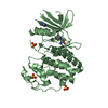
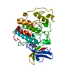
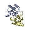
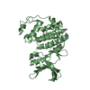
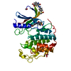
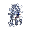
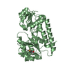
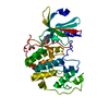
- PDB-7lm5: Crystal structure of the Zn(II)-bound AdcAII H65A mutant variant ... -
+ Open data

- Basic information
| Entry | Database: PDB / ID: 7lm5 | ||||||
|---|---|---|---|---|---|---|---|
| Title | Crystal structure of the Zn(II)-bound AdcAII H65A mutant variant of Streptococcus pneumoniae | ||||||
 Components Components | Adhesion protein | ||||||
 Keywords Keywords | METAL BINDING PROTEIN / SBP / ATP-binding cassette transporter / Zn acquisition | ||||||
| Function / homology | Adhesin B / Adhesion lipoprotein / Periplasmic solute binding protein, ZnuA-like / Zinc-uptake complex component A periplasmic / metal ion transport / Prokaryotic membrane lipoprotein lipid attachment site profile. / cell adhesion / metal ion binding / Adhesion protein Function and homology information Function and homology information | ||||||
| Biological species |  Streptococcus pseudopneumoniae 5247 (bacteria) Streptococcus pseudopneumoniae 5247 (bacteria) | ||||||
| Method |  X-RAY DIFFRACTION / SYNCHROTRON / MOLECULAR REPLACEMENT / Resolution: 2.4 Å X-RAY DIFFRACTION / SYNCHROTRON / MOLECULAR REPLACEMENT / Resolution: 2.4 Å | ||||||
 Authors Authors | Luo, Z. / Zupan, M. / McDevitt, C.A. / Kobe, B. | ||||||
| Funding support |  Australia, 1items Australia, 1items
| ||||||
 Citation Citation | Journal: Front Cell Infect Microbiol / Year: 2021 Title: Conformation of the Solute-Binding Protein AdcAII Influences Zinc Uptake in Streptococcus pneumoniae . Authors: Zupan, M.L. / Luo, Z. / Ganio, K. / Pederick, V.G. / Neville, S.L. / Deplazes, E. / Kobe, B. / McDevitt, C.A. | ||||||
| History |
|
- Structure visualization
| Structure viewer | Molecule: MolmilJmol/JSmol |
|---|
- Downloads & links
-Download
| PDBx/mmCIF format | 7lm5.cif.gz | 129.4 KB | Display | PDBx/mmCIF format |
|---|---|---|---|---|
| PDB format | pdb7lm5.ent.gz | 93.6 KB | Display | PDB format |
| PDBx/mmJSON format | 7lm5.json.gz | Tree view | PDBx/mmJSON format | |
| Others |  Other downloads Other downloads |
-Validation report
| Arichive directory | https://data.pdbj.org/pub/pdb/validation_reports/lm/7lm5ftp://data.pdbj.org/pub/pdb/validation_reports/lm/7lm5 | HTTPS FTP |
|---|
-Related structure data
| Related structure data |  7lm6C  7lm7C  3cx3S S: Starting model for refinement C: citing same article ( |
|---|---|
| Similar structure data |


-Links
 PDBj
PDBj


- Assembly
| Deposited unit | 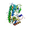
| ||||||||||||
|---|---|---|---|---|---|---|---|---|---|---|---|---|---|
| 1 |
| ||||||||||||
| Unit cell |
| ||||||||||||
| Components on special symmetry positions |
|
-Components
| #1: Protein | Mass: 32650.613 Da / Num. of mol.: 1 / Mutation: H65A Source method: isolated from a genetically manipulated source Source: (gene. exp.) Streptococcus pseudopneumoniae 5247 (bacteria)Gene: U753_04975 Production host: References: UniProt: V8IJK5 | ||||||||
|---|---|---|---|---|---|---|---|---|---|
| #2: Chemical | ChemComp-ZN /   Mass: 65.409 Da / Num. of mol.: 11 / Source method: obtained synthetically / Formula: Zn / Feature type: SUBJECT OF INVESTIGATION Mass: 65.409 Da / Num. of mol.: 11 / Source method: obtained synthetically / Formula: Zn / Feature type: SUBJECT OF INVESTIGATION#3: Chemical |   Mass: 35.453 Da / Num. of mol.: 2 / Source method: obtained synthetically / Formula: Cl Mass: 35.453 Da / Num. of mol.: 2 / Source method: obtained synthetically / Formula: Cl#4: Chemical | ChemComp-NA / |   Mass: 22.990 Da / Num. of mol.: 1 / Source method: obtained synthetically / Formula: Na Mass: 22.990 Da / Num. of mol.: 1 / Source method: obtained synthetically / Formula: Na#5: Water | ChemComp-HOH / |  Mass: 18.015 Da / Num. of mol.: 60 / Source method: isolated from a natural source / Formula: H2O Mass: 18.015 Da / Num. of mol.: 60 / Source method: isolated from a natural source / Formula: H2OHas ligand of interest | Y | |
-Experimental details
-Experiment
| Experiment | Method: X-RAY DIFFRACTION / Number of used crystals: 1 |
|---|
- Sample preparation
| Crystal | Density Matthews: 2.27 Å3/Da / Density % sol: 40.8 % |
|---|---|
| Crystal grow | Temperature: 293 K / Method: vapor diffusion, hanging drop / pH: 8 Details: 0.1 M imidazole pH 8.0, 0.2 M zinc acetate, 20% (w/v) polyethylene glycol 3350 |
-Data collection
| Diffraction | Mean temperature: 100 K / Serial crystal experiment: N |
|---|---|
| Diffraction source | Source: SYNCHROTRON / Site: Australian Synchrotron / Beamline: MX2 / Wavelength: 0.954 Å |
| Detector | Type: DECTRIS EIGER X 16M / Detector: PIXEL / Date: May 15, 2017 |
| Radiation | Protocol: SINGLE WAVELENGTH / Monochromatic (M) / Laue (L): M / Scattering type: x-ray |
| Radiation wavelength | Wavelength: 0.954 Å / Relative weight: 1 |
| Reflection | Resolution: 2.4→36.16 Å / Num. obs: 10512 / % possible obs: 99.6 % / Redundancy: 7.5 % / Biso Wilson estimate: 38.25 Å2 / CC1/2: 0.998 / Net I/σ(I): 16.9 |
| Reflection shell | Resolution: 2.4→2.49 Å / Rmerge(I) obs: 0.298 / Num. unique obs: 1013 / CC1/2: 0.871 |
- Processing
| Software |
| |||||||||||||||||||||||||||||||||||
|---|---|---|---|---|---|---|---|---|---|---|---|---|---|---|---|---|---|---|---|---|---|---|---|---|---|---|---|---|---|---|---|---|---|---|---|---|
| Refinement | Method to determine structure: MOLECULAR REPLACEMENT Starting model: 3CX3 Resolution: 2.4→36.16 Å / SU ML: 0.2167 / Cross valid method: FREE R-VALUE / σ(F): 1.35 / Phase error: 29.0247 / Stereochemistry target values: CDL v1.2
| |||||||||||||||||||||||||||||||||||
| Solvent computation | Shrinkage radii: 0.9 Å / VDW probe radii: 1.11 Å / Solvent model: FLAT BULK SOLVENT MODEL | |||||||||||||||||||||||||||||||||||
| Displacement parameters | Biso mean: 51.5 Å2 | |||||||||||||||||||||||||||||||||||
| Refinement step | Cycle: LAST / Resolution: 2.4→36.16 Å
| |||||||||||||||||||||||||||||||||||
| Refine LS restraints |
| |||||||||||||||||||||||||||||||||||
| LS refinement shell |
|
