 Movie
Movie Controller


+ Open data

- Basic information
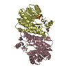
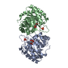
| Entry | Database: PDB / ID: 7lg9 | ||||||
|---|---|---|---|---|---|---|---|
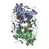
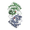
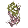
| Title | ChsB1 | ||||||
 Components Components | 3-ketoacyl-ACP reductase | ||||||
 Keywords Keywords | OXIDOREDUCTASE / short-chain type alcohol dehydrogenase/reductase hydroxyacyl-CoA dehydrogenase | ||||||
| Function / homology |  Function and homology information Function and homology information3-oxoacyl-[acyl-carrier-protein] reductase / 3-oxoacyl-[acyl-carrier-protein] reductase (NADPH) activity / Oxidoreductases; Acting on the CH-OH group of donors; With NAD+ or NADP+ as acceptor Similarity search - Function | ||||||
| Biological species |   Mycobacterium tuberculosis (bacteria) Mycobacterium tuberculosis (bacteria) | ||||||
| Method |  X-RAY DIFFRACTION / SYNCHROTRON / MOLECULAR REPLACEMENT / Resolution: 2.03 Å X-RAY DIFFRACTION / SYNCHROTRON / MOLECULAR REPLACEMENT / Resolution: 2.03 Å | ||||||
 Authors Authors | Yuan, T. / Werman, J.M. / Yin, X. / Yang, M. / Garcia-Diaz, M. / Sampson, N.S. | ||||||
| Funding support |  United States, 1items United States, 1items
| ||||||
 Citation Citation | Journal: Acs Infect Dis. / Year: 2021 Title: Enzymatic beta-Oxidation of the Cholesterol Side Chain in Mycobacterium tuberculosis Bifurcates Stereospecifically at Hydration of 3-Oxo-cholest-4,22-dien-24-oyl-CoA. Authors: Yuan, T. / Werman, J.M. / Yin, X. / Yang, M. / Garcia-Diaz, M. / Sampson, N.S. | ||||||
| History |
|
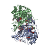
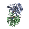
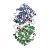
- Structure visualization
| Structure viewer | Molecule: MolmilJmol/JSmol |
|---|
- Downloads & links
-Download
| PDBx/mmCIF format | 7lg9.cif.gz | 121.3 KB | Display | PDBx/mmCIF format |
|---|---|---|---|---|
| PDB format | pdb7lg9.ent.gz | 84.6 KB | Display | PDB format |
| PDBx/mmJSON format | 7lg9.json.gz | Tree view | PDBx/mmJSON format | |
| Others |  Other downloads Other downloads |
-Validation report
| Arichive directory | https://data.pdbj.org/pub/pdb/validation_reports/lg/7lg9ftp://data.pdbj.org/pub/pdb/validation_reports/lg/7lg9 | HTTPS FTP |
|---|
-Related structure data
| Related structure data | 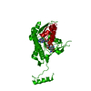 7lgbC  4kzpS S: Starting model for refinement C: citing same article ( |
|---|---|
| Similar structure data |

-Links
 PDBj
PDBj


- Assembly
| Deposited unit | 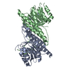
| ||||||||||||
|---|---|---|---|---|---|---|---|---|---|---|---|---|---|
| 1 |
| ||||||||||||
| Unit cell |
|
-Components
| #1: Protein | Mass: 32764.697 Da / Num. of mol.: 2 Source method: isolated from a genetically manipulated source Source: (gene. exp.) Mycobacterium tuberculosis (bacteria)Gene: fabG_8, fabG_1, fabG_2, fabG_4, fabG_6, fabG_7, DSI38_08445, E5M52_16205, ERS007665_00489, ERS007670_00458, ERS007679_00994, ERS007681_00640, ERS007741_01258, ERS013471_03025, ERS023446_03243, ...Gene: fabG_8, fabG_1, fabG_2, fabG_4, fabG_6, fabG_7, DSI38_08445, E5M52_16205, ERS007665_00489, ERS007670_00458, ERS007679_00994, ERS007681_00640, ERS007741_01258, ERS013471_03025, ERS023446_03243, ERS024276_00059, ERS075361_03673, F6W99_02176, FRD82_16680, SAMEA2683035_03264 Production host: Rhodococcus jostii RHA1 (bacteria)References: UniProt: A0A045J1S8, Oxidoreductases; Acting on the CH-OH group of donors; With NAD+ or NADP+ as acceptor, 3-oxoacyl-[acyl-carrier-protein] reductase #2: Water | ChemComp-HOH / |  Mass: 18.015 Da / Num. of mol.: 108 / Source method: isolated from a natural source / Formula: H2O Mass: 18.015 Da / Num. of mol.: 108 / Source method: isolated from a natural source / Formula: H2O |
|---|
-Experimental details
-Experiment
| Experiment | Method: X-RAY DIFFRACTION / Number of used crystals: 1 |
|---|
- Sample preparation
| Crystal | Density Matthews: 2.24 Å3/Da / Density % sol: 45.15 % |
|---|---|
| Crystal grow | Temperature: 297.15 K / Method: vapor diffusion, hanging drop Details: 15% w/v PEG4000, 0.2M ammonium acetate, 0.1M sodium citrate pH5.6 |
-Data collection
| Diffraction | Mean temperature: 80 K / Serial crystal experiment: N |
|---|---|
| Diffraction source | Source: SYNCHROTRON / Site: NSLS-II / Beamline: 17-ID-1 / Wavelength: 0.979 Å |
| Detector | Type: DECTRIS EIGER2 X 9M / Detector: PIXEL / Date: Feb 21, 2020 |
| Radiation | Protocol: SINGLE WAVELENGTH / Monochromatic (M) / Laue (L): M / Scattering type: x-ray |
| Radiation wavelength | Wavelength: 0.979 Å / Relative weight: 1 |
| Reflection | Resolution: 2.03→81.77 Å / Num. obs: 122908 / % possible obs: 93.1 % / Redundancy: 5.5 % / Biso Wilson estimate: 30.45 Å2 / CC1/2: 0.991 / Rmerge(I) obs: 0.2 / Net I/σ(I): 6.8 |
| Reflection shell | Resolution: 2.03→2.31 Å / Rmerge(I) obs: 1.13 / Num. unique obs: 122908 / CC1/2: 0.665 |
- Processing
| Software |
| |||||||||||||||||||||||||||||||||||||||||||||||||||||||||||||||||||||||||||||||||||||||||||||||||||||||||||||||||||||||
|---|---|---|---|---|---|---|---|---|---|---|---|---|---|---|---|---|---|---|---|---|---|---|---|---|---|---|---|---|---|---|---|---|---|---|---|---|---|---|---|---|---|---|---|---|---|---|---|---|---|---|---|---|---|---|---|---|---|---|---|---|---|---|---|---|---|---|---|---|---|---|---|---|---|---|---|---|---|---|---|---|---|---|---|---|---|---|---|---|---|---|---|---|---|---|---|---|---|---|---|---|---|---|---|---|---|---|---|---|---|---|---|---|---|---|---|---|---|---|---|---|
| Refinement | Method to determine structure: MOLECULAR REPLACEMENT Starting model: 4KZP Resolution: 2.03→81.77 Å / SU ML: 0.2619 / Cross valid method: FREE R-VALUE / σ(F): 1.35 / Phase error: 31.5391 Stereochemistry target values: GeoStd + Monomer Library + CDL v1.2
| |||||||||||||||||||||||||||||||||||||||||||||||||||||||||||||||||||||||||||||||||||||||||||||||||||||||||||||||||||||||
| Solvent computation | Shrinkage radii: 0.9 Å / VDW probe radii: 1.11 Å / Solvent model: FLAT BULK SOLVENT MODEL | |||||||||||||||||||||||||||||||||||||||||||||||||||||||||||||||||||||||||||||||||||||||||||||||||||||||||||||||||||||||
| Displacement parameters | Biso mean: 36.91 Å2 | |||||||||||||||||||||||||||||||||||||||||||||||||||||||||||||||||||||||||||||||||||||||||||||||||||||||||||||||||||||||
| Refinement step | Cycle: LAST / Resolution: 2.03→81.77 Å
| |||||||||||||||||||||||||||||||||||||||||||||||||||||||||||||||||||||||||||||||||||||||||||||||||||||||||||||||||||||||
| Refine LS restraints |
| |||||||||||||||||||||||||||||||||||||||||||||||||||||||||||||||||||||||||||||||||||||||||||||||||||||||||||||||||||||||
| LS refinement shell |
|
