[English] 日本語
 Yorodumi
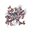
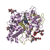
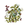
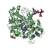
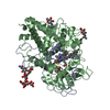
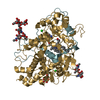
- PDB-7lan: CRYSTAL STRUCTURE OF MYELOPEROXIDASE SUBFORM C (MPO) COMPLEX WITH... -
+ Open data
- Basic information
| Entry | Database: PDB / ID: 7lan | ||||||
|---|---|---|---|---|---|---|---|
| Title | CRYSTAL STRUCTURE OF MYELOPEROXIDASE SUBFORM C (MPO) COMPLEX WITH COMPOUND-30 AKA 7-[(3~{S},4~{R},6~{R})-4-benzyl-2-oxa-7,13,14-triazatetracyclo[14.3.1.1^{3,6}.1^{11,14}]docosa-1(19),11(21),12,16(20),17-pentaen-10-yl]-3~{H}-triazolo[4,5-b]pyridin-5-amine | ||||||
 Components Components |
| ||||||
 Keywords Keywords | OXIDOREDUCTASE / MYELOPEROXIDASE | ||||||
| Function / homology |  Function and homology information Function and homology informationmyeloperoxidase / hypochlorous acid biosynthetic process / Events associated with phagocytolytic activity of PMN cells / phagocytic vesicle lumen / response to gold nanoparticle / response to yeast / respiratory burst involved in defense response / low-density lipoprotein particle remodeling / azurophil granule / response to food ...myeloperoxidase / hypochlorous acid biosynthetic process / Events associated with phagocytolytic activity of PMN cells / phagocytic vesicle lumen / response to gold nanoparticle / response to yeast / respiratory burst involved in defense response / low-density lipoprotein particle remodeling / azurophil granule / response to food / defense response to fungus / response to mechanical stimulus / removal of superoxide radicals / secretory granule / hydrogen peroxide catabolic process / peroxidase activity / defense response / azurophil granule lumen / heparin binding / response to oxidative stress / response to lipopolysaccharide / lysosome / defense response to bacterium / intracellular membrane-bounded organelle / heme binding / Neutrophil degranulation / chromatin binding / negative regulation of apoptotic process / extracellular space / extracellular exosome / extracellular region / nucleoplasm / metal ion binding / nucleus Similarity search - Function | ||||||
| Biological species |  Homo sapiens (human) Homo sapiens (human) | ||||||
| Method |  X-RAY DIFFRACTION / X-RAY DIFFRACTION /  SYNCHROTRON / SYNCHROTRON /  MOLECULAR REPLACEMENT / Resolution: 2.28 Å MOLECULAR REPLACEMENT / Resolution: 2.28 Å | ||||||
 Authors Authors | Khan, J.A. | ||||||
 Citation Citation |  Journal: Bioorg.Med.Chem.Lett. / Year: 2021 Journal: Bioorg.Med.Chem.Lett. / Year: 2021Title: Small molecule and macrocyclic pyrazole derived inhibitors of myeloperoxidase (MPO). Authors: Hu, C.H. / Neissel Valente, M.W. / Halpern, O.S. / Jusuf, S. / Khan, J.A. / Locke, G.A. / Duke, G.J. / Liu, X. / Duclos, F.J. / Wexler, R.R. / Kick, E.K. / Smallheer, J.M. | ||||||
| History |
|
- Structure visualization
| Structure viewer | Molecule:  Molmil Molmil Jmol/JSmol Jmol/JSmol |
|---|
- Downloads & links
- Download
| PDBx/mmCIF format |  7lan.cif.gz 7lan.cif.gz | 495.1 KB | Display |  PDBx/mmCIF format PDBx/mmCIF format |
|---|---|---|---|---|
| PDB format |  pdb7lan.ent.gz pdb7lan.ent.gz | 403.4 KB | Display |  PDB format PDB format |
| PDBx/mmJSON format |  7lan.json.gz 7lan.json.gz | Tree view |  PDBx/mmJSON format PDBx/mmJSON format | |
| Others |  Other downloads Other downloads |
-Validation report
| Summary document |  7lan_validation.pdf.gz 7lan_validation.pdf.gz | 5.2 MB | Display |  wwPDB validaton report wwPDB validaton report |
|---|---|---|---|---|
| Full document |  7lan_full_validation.pdf.gz 7lan_full_validation.pdf.gz | 5.1 MB | Display | |
| Data in XML |  7lan_validation.xml.gz 7lan_validation.xml.gz | 91.6 KB | Display | |
| Data in CIF |  7lan_validation.cif.gz 7lan_validation.cif.gz | 128.1 KB | Display | |
| Arichive directory |  https://data.pdbj.org/pub/pdb/validation_reports/la/7lan https://data.pdbj.org/pub/pdb/validation_reports/la/7lan ftp://data.pdbj.org/pub/pdb/validation_reports/la/7lan ftp://data.pdbj.org/pub/pdb/validation_reports/la/7lan | HTTPS FTP |
-Related structure data
| Related structure data |  7laeC  7lagC  7lalC  6wy7S S: Starting model for refinement C: citing same article ( |
|---|---|
| Similar structure data |
- Links
Links
- Assembly
| Deposited unit | 
| ||||||||
|---|---|---|---|---|---|---|---|---|---|
| 1 | 
| ||||||||
| 2 | 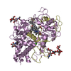
| ||||||||
| 3 | 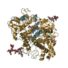
| ||||||||
| 4 | 
| ||||||||
| Unit cell |
|
- Components
-Protein , 2 types, 8 molecules ADFHBEGI
| #1: Protein | Mass: 11974.420 Da / Num. of mol.: 4 / Source method: isolated from a natural source / Details: Blood Neutrophill / Source: (natural)  Homo sapiens (human) / References: UniProt: P05164, myeloperoxidase Homo sapiens (human) / References: UniProt: P05164, myeloperoxidase#2: Protein | Mass: 53218.188 Da / Num. of mol.: 4 / Source method: isolated from a natural source / Source: (natural)  Homo sapiens (human) / References: UniProt: P05164, myeloperoxidase Homo sapiens (human) / References: UniProt: P05164, myeloperoxidase |
|---|
-Sugars , 3 types, 12 molecules 
| #3: Polysaccharide | Source method: isolated from a genetically manipulated source #4: Polysaccharide | alpha-D-mannopyranose-(1-3)-[alpha-D-mannopyranose-(1-6)]beta-D-mannopyranose-(1-4)-2-acetamido-2- ...alpha-D-mannopyranose-(1-3)-[alpha-D-mannopyranose-(1-6)]beta-D-mannopyranose-(1-4)-2-acetamido-2-deoxy-beta-D-glucopyranose-(1-4)-[alpha-L-fucopyranose-(1-6)]2-acetamido-2-deoxy-beta-D-glucopyranose Source method: isolated from a genetically manipulated source #7: Sugar | ChemComp-NAG / |
|---|
-Non-polymers , 5 types, 982 molecules 


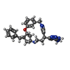


| #5: Chemical | ChemComp-CL / #6: Chemical | ChemComp-HEM / #8: Chemical | ChemComp-CA / #9: Chemical | ChemComp-XS1 / #10: Water | ChemComp-HOH / | |
|---|
-Details
| Has ligand of interest | Y |
|---|---|
| Has protein modification | Y |
-Experimental details
-Experiment
| Experiment | Method:  X-RAY DIFFRACTION / Number of used crystals: 1 X-RAY DIFFRACTION / Number of used crystals: 1 |
|---|
- Sample preparation
| Crystal | Density Matthews: 2.4 Å3/Da / Density % sol: 48.78 % |
|---|---|
| Crystal grow | Temperature: 296 K / Method: vapor diffusion, hanging drop Details: 0.1M Hepes pH 7.5, 150mM NaCl, 20-25%(V/V)PEG3350.Crystals were cryoprotected by supplementing the mother liquor with 15% (v/v) ethylene glycol and harvested by flash-cooling in liquid nitrogen PH range: 7.5? |
-Data collection
| Diffraction | Mean temperature: 100 K / Serial crystal experiment: N |
|---|---|
| Diffraction source | Source:  SYNCHROTRON / Site: SYNCHROTRON / Site:  CLSI CLSI  / Beamline: 08ID-1 / Wavelength: 1 Å / Beamline: 08ID-1 / Wavelength: 1 Å |
| Detector | Type: RAYONIX MX-300 / Detector: CCD / Date: Sep 6, 2014 |
| Radiation | Protocol: SINGLE WAVELENGTH / Monochromatic (M) / Laue (L): M / Scattering type: x-ray |
| Radiation wavelength | Wavelength: 1 Å / Relative weight: 1 |
| Reflection | Resolution: 2.28→50 Å / Num. obs: 114459 / % possible obs: 100 % / Redundancy: 6.3 % / CC1/2: 0.8 / Net I/σ(I): 18.7 |
| Reflection shell | Resolution: 2.28→2.36 Å / Rmerge(I) obs: 0.72 / Num. unique obs: 11351 |
- Processing
| Software |
| ||||||||||||||||||||||||||||||||||||||||||||||||||||||||||||||||||||||||||||||||||||||||||||||||||||||||||||
|---|---|---|---|---|---|---|---|---|---|---|---|---|---|---|---|---|---|---|---|---|---|---|---|---|---|---|---|---|---|---|---|---|---|---|---|---|---|---|---|---|---|---|---|---|---|---|---|---|---|---|---|---|---|---|---|---|---|---|---|---|---|---|---|---|---|---|---|---|---|---|---|---|---|---|---|---|---|---|---|---|---|---|---|---|---|---|---|---|---|---|---|---|---|---|---|---|---|---|---|---|---|---|---|---|---|---|---|---|---|
| Refinement | Method to determine structure:  MOLECULAR REPLACEMENT MOLECULAR REPLACEMENTStarting model: 6WY7 Resolution: 2.28→47.45 Å / Cor.coef. Fo:Fc: 0.945 / Cor.coef. Fo:Fc free: 0.9253 / SU R Cruickshank DPI: 0.34 / Cross valid method: THROUGHOUT / σ(F): 0 / SU R Blow DPI: 0.343 / SU Rfree Blow DPI: 0.224 / SU Rfree Cruickshank DPI: 0.226
| ||||||||||||||||||||||||||||||||||||||||||||||||||||||||||||||||||||||||||||||||||||||||||||||||||||||||||||
| Displacement parameters | Biso max: 117.94 Å2 / Biso mean: 44.53 Å2 / Biso min: 5.91 Å2
| ||||||||||||||||||||||||||||||||||||||||||||||||||||||||||||||||||||||||||||||||||||||||||||||||||||||||||||
| Refine analyze | Luzzati coordinate error obs: 0.293 Å | ||||||||||||||||||||||||||||||||||||||||||||||||||||||||||||||||||||||||||||||||||||||||||||||||||||||||||||
| Refinement step | Cycle: final / Resolution: 2.28→47.45 Å
| ||||||||||||||||||||||||||||||||||||||||||||||||||||||||||||||||||||||||||||||||||||||||||||||||||||||||||||
| Refine LS restraints |
| ||||||||||||||||||||||||||||||||||||||||||||||||||||||||||||||||||||||||||||||||||||||||||||||||||||||||||||
| LS refinement shell | Resolution: 2.28→2.34 Å / Rfactor Rfree error: 0 / Total num. of bins used: 20
|
 Movie
Movie Controller




 PDBj
PDBj


