[English] 日本語
 Yorodumi
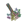
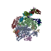
- PDB-7kxr: Protective antigen pore translocating lethal factor N-terminal domain -
+ Open data
- Basic information
| Entry | Database: PDB / ID: 7kxr | ||||||
|---|---|---|---|---|---|---|---|
| Title | Protective antigen pore translocating lethal factor N-terminal domain | ||||||
 Components Components |
| ||||||
 Keywords Keywords | TOXIN / translocation / complex / anthrax / refolding | ||||||
| Function / homology |  Function and homology information Function and homology informationanthrax lethal factor endopeptidase / symbiont-mediated suppression of host MAPK cascade / host cell cytosol / Uptake and function of anthrax toxins / host cell endosome membrane / metalloendopeptidase activity / protein homooligomerization / metallopeptidase activity / toxin activity / host cell plasma membrane ...anthrax lethal factor endopeptidase / symbiont-mediated suppression of host MAPK cascade / host cell cytosol / Uptake and function of anthrax toxins / host cell endosome membrane / metalloendopeptidase activity / protein homooligomerization / metallopeptidase activity / toxin activity / host cell plasma membrane / proteolysis / extracellular region / zinc ion binding / metal ion binding / identical protein binding / membrane Similarity search - Function | ||||||
| Biological species |  | ||||||
| Method | ELECTRON MICROSCOPY / single particle reconstruction / cryo EM / Resolution: 3.3 Å | ||||||
 Authors Authors | Machen, A.J. / Freudenthal, B.D. | ||||||
| Funding support |  United States, 1items United States, 1items
| ||||||
 Citation Citation |  Journal: Sci Rep / Year: 2021 Journal: Sci Rep / Year: 2021Title: Anthrax toxin translocation complex reveals insight into the lethal factor unfolding and refolding mechanism. Authors: Alexandra J Machen / Mark T Fisher / Bret D Freudenthal /  Abstract: Translocation is essential to the anthrax toxin mechanism. Protective antigen (PA), the binding component of this AB toxin, forms an oligomeric pore that translocates lethal factor (LF) or edema ...Translocation is essential to the anthrax toxin mechanism. Protective antigen (PA), the binding component of this AB toxin, forms an oligomeric pore that translocates lethal factor (LF) or edema factor, the active components of the toxin, into the cell. Structural details of the translocation process have remained elusive despite their biological importance. To overcome the technical challenges of studying translocation intermediates, we developed a method to immobilize, transition, and stabilize anthrax toxin to mimic important physiological steps in the intoxication process. Here, we report a cryoEM snapshot of PA translocating the N-terminal domain of LF (LF). The resulting 3.3 Å structure of the complex shows density of partially unfolded LF near the canonical PA binding site. Interestingly, we also observe density consistent with an α helix emerging from the 100 Å β barrel channel suggesting LF secondary structural elements begin to refold in the pore channel. We conclude the anthrax toxin β barrel aids in efficient folding of its enzymatic payload prior to channel exit. Our hypothesized refolding mechanism has broader implications for pore length of other protein translocating toxins. | ||||||
| History |
|
- Structure visualization
| Movie |
 Movie viewer Movie viewer |
|---|---|
| Structure viewer | Molecule:  Molmil Molmil Jmol/JSmol Jmol/JSmol |
- Downloads & links
- Download
| PDBx/mmCIF format |  7kxr.cif.gz 7kxr.cif.gz | 700.5 KB | Display |  PDBx/mmCIF format PDBx/mmCIF format |
|---|---|---|---|---|
| PDB format |  pdb7kxr.ent.gz pdb7kxr.ent.gz | 588.5 KB | Display |  PDB format PDB format |
| PDBx/mmJSON format |  7kxr.json.gz 7kxr.json.gz | Tree view |  PDBx/mmJSON format PDBx/mmJSON format | |
| Others |  Other downloads Other downloads |
-Validation report
| Arichive directory |  https://data.pdbj.org/pub/pdb/validation_reports/kx/7kxr https://data.pdbj.org/pub/pdb/validation_reports/kx/7kxr ftp://data.pdbj.org/pub/pdb/validation_reports/kx/7kxr ftp://data.pdbj.org/pub/pdb/validation_reports/kx/7kxr | HTTPS FTP |
|---|
-Related structure data
| Related structure data |  23066MC M: map data used to model this data C: citing same article ( |
|---|---|
| Similar structure data |
- Links
Links
- Assembly
| Deposited unit | 
|
|---|---|
| 1 |
|
- Components
| #1: Protein | Mass: 30520.408 Da / Num. of mol.: 1 / Fragment: UNP Residues 34-296 / Mutation: E126C Source method: isolated from a genetically manipulated source Source: (gene. exp.)   References: UniProt: P15917, anthrax lethal factor endopeptidase | ||||
|---|---|---|---|---|---|
| #2: Protein | Mass: 63019.012 Da / Num. of mol.: 7 / Fragment: UNP Residues 203-764 Source method: isolated from a genetically manipulated source Source: (gene. exp.)   #3: Chemical | ChemComp-CA / Has ligand of interest | N | |
-Experimental details
-Experiment
| Experiment | Method: ELECTRON MICROSCOPY |
|---|---|
| EM experiment | Aggregation state: PARTICLE / 3D reconstruction method: single particle reconstruction |
- Sample preparation
| Component | Name: Anthrax toxin protective antigen translocating lethal factor N-terminal domain Type: COMPLEX / Entity ID: #1-#2 / Source: RECOMBINANT |
|---|---|
| Source (natural) | Organism:  |
| Source (recombinant) | Organism:  |
| Buffer solution | pH: 5.5 |
| Specimen | Embedding applied: NO / Shadowing applied: NO / Staining applied: NO / Vitrification applied: YES |
| Vitrification | Instrument: FEI VITROBOT MARK IV / Cryogen name: ETHANE |
- Electron microscopy imaging
| Experimental equipment |  Model: Titan Krios / Image courtesy: FEI Company |
|---|---|
| Microscopy | Model: FEI TITAN KRIOS |
| Electron gun | Electron source:  FIELD EMISSION GUN / Accelerating voltage: 300 kV / Illumination mode: FLOOD BEAM FIELD EMISSION GUN / Accelerating voltage: 300 kV / Illumination mode: FLOOD BEAM |
| Electron lens | Mode: BRIGHT FIELD |
| Image recording | Electron dose: 40 e/Å2 / Detector mode: SUPER-RESOLUTION / Film or detector model: GATAN K2 SUMMIT (4k x 4k) |
- Processing
| EM software |
| ||||||||||||
|---|---|---|---|---|---|---|---|---|---|---|---|---|---|
| CTF correction | Type: PHASE FLIPPING AND AMPLITUDE CORRECTION | ||||||||||||
| 3D reconstruction | Resolution: 3.3 Å / Resolution method: FSC 0.143 CUT-OFF / Num. of particles: 122651 / Symmetry type: POINT |
 Movie
Movie Controller


 PDBj
PDBj


