 Movie
Movie Controller

[English] 日本語
 Yorodumi
- PDB-7kur: Crystal Structure of Danio rerio Histone Deacetylase 10 Y307F Mut... -
+ Open data

- Basic information
| Entry | Database: PDB / ID: 7kur | ||||||
|---|---|---|---|---|---|---|---|
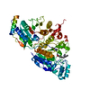
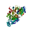
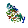
| Title | Crystal Structure of Danio rerio Histone Deacetylase 10 Y307F Mutant in Complex with N-Acetylputrescine | ||||||
 Components Components | Polyamine deacetylase HDAC10 | ||||||
 Keywords Keywords | HYDROLASE / Histone Deacetylase | ||||||
| Function / homology |  Function and homology information Function and homology information: / : / HDACs deacetylate histones / acetylspermidine deacetylase / acetylspermidine deacetylase activity / acetylputrescine deacetylase / acetylputrescine deacetylase activity / deacetylase activity / homologous recombination / swimming behavior ...: / : / HDACs deacetylate histones / acetylspermidine deacetylase / acetylspermidine deacetylase activity / acetylputrescine deacetylase / acetylputrescine deacetylase activity / deacetylase activity / homologous recombination / swimming behavior / macroautophagy / zinc ion binding / nucleus / cytoplasm / cytosol Similarity search - Function | ||||||
| Biological species |  | ||||||
| Method |  X-RAY DIFFRACTION / SYNCHROTRON / MOLECULAR REPLACEMENT / Resolution: 2.1 Å X-RAY DIFFRACTION / SYNCHROTRON / MOLECULAR REPLACEMENT / Resolution: 2.1 Å | ||||||
 Authors Authors | Herbst-Gervasoni, C.J. / Christianson, D.W. | ||||||
| Funding support |  United States, 1items United States, 1items
| ||||||
 Citation Citation | Journal: Biochemistry / Year: 2021 Title: X-ray Crystallographic Snapshots of Substrate Binding in the Active Site of Histone Deacetylase 10. Authors: Herbst-Gervasoni, C.J. / Christianson, D.W. | ||||||
| History |
|
- Structure visualization
| Structure viewer | Molecule: MolmilJmol/JSmol |
|---|
- Downloads & links
-Download
| PDBx/mmCIF format | 7kur.cif.gz | 175.8 KB | Display | PDBx/mmCIF format |
|---|---|---|---|---|
| PDB format | pdb7kur.ent.gz | 108.3 KB | Display | PDB format |
| PDBx/mmJSON format | 7kur.json.gz | Tree view | PDBx/mmJSON format | |
| Others |  Other downloads Other downloads |
-Validation report
| Arichive directory | https://data.pdbj.org/pub/pdb/validation_reports/ku/7kurftp://data.pdbj.org/pub/pdb/validation_reports/ku/7kur | HTTPS FTP |
|---|
-Related structure data
| Related structure data | 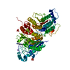 7kuqC  7kusC  7kutC  7kuvC 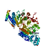 5td7S S: Starting model for refinement C: citing same article ( |
|---|---|
| Similar structure data |


-Links
 PDBj
PDBj- Assembly
| Deposited unit | 
| ||||||||||||
|---|---|---|---|---|---|---|---|---|---|---|---|---|---|
| 1 |
| ||||||||||||
| Unit cell |
|
-Components
-Protein , 1 types, 1 molecules A
| #1: Protein | Mass: 75240.820 Da / Num. of mol.: 1 / Mutation: Y307F Source method: isolated from a genetically manipulated source Source: (gene. exp.)  References: UniProt: F1QCV2, acetylspermidine deacetylase, acetylputrescine deacetylase |
|---|
-Non-polymers , 7 types, 265 molecules 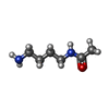


| #2: Chemical | ChemComp-X5A /  Mass: 130.188 Da / Num. of mol.: 1 / Source method: obtained synthetically / Formula: C6H14N2O / Feature type: SUBJECT OF INVESTIGATION Mass: 130.188 Da / Num. of mol.: 1 / Source method: obtained synthetically / Formula: C6H14N2O / Feature type: SUBJECT OF INVESTIGATION | ||||||
|---|---|---|---|---|---|---|---|
| #3: Chemical | ChemComp-PEG /  Mass: 106.120 Da / Num. of mol.: 1 / Source method: obtained synthetically / Formula: C4H10O3 Mass: 106.120 Da / Num. of mol.: 1 / Source method: obtained synthetically / Formula: C4H10O3 | ||||||
| #4: Chemical | ChemComp-ZN /  Mass: 65.409 Da / Num. of mol.: 1 / Source method: obtained synthetically / Formula: Zn Mass: 65.409 Da / Num. of mol.: 1 / Source method: obtained synthetically / Formula: Zn | ||||||
| #5: Chemical |  Mass: 39.098 Da / Num. of mol.: 2 / Source method: obtained synthetically / Formula: K Mass: 39.098 Da / Num. of mol.: 2 / Source method: obtained synthetically / Formula: K#6: Chemical |  Mass: 94.971 Da / Num. of mol.: 3 / Source method: obtained synthetically / Formula: PO4 Mass: 94.971 Da / Num. of mol.: 3 / Source method: obtained synthetically / Formula: PO4#7: Chemical | ChemComp-NA / |  Mass: 22.990 Da / Num. of mol.: 1 / Source method: obtained synthetically / Formula: Na Mass: 22.990 Da / Num. of mol.: 1 / Source method: obtained synthetically / Formula: Na#8: Water | ChemComp-HOH / | Mass: 18.015 Da / Num. of mol.: 256 / Source method: isolated from a natural source / Formula: H2O |
-Details
| Has ligand of interest | Y |
|---|
-Experimental details
-Experiment
| Experiment | Method: X-RAY DIFFRACTION / Number of used crystals: 1 |
|---|
- Sample preparation
| Crystal | Density Matthews: 3.06 Å3/Da / Density % sol: 59.79 % |
|---|---|
| Crystal grow | Temperature: 277 K / Method: vapor diffusion, sitting drop Details: 10 mg/mL HDAC10, 10 mM N-Acetylputrescine, 1:1000 trypsin:HDAC10, 0.100 M sodium phosphate monobasic, 0.100 M sodium phosphate dibasic, 20% PEG3350 (w/v), HDAC10 microseed |
-Data collection
| Diffraction | Mean temperature: 100 K / Serial crystal experiment: N |
|---|---|
| Diffraction source | Source: SYNCHROTRON / Site: APS / Beamline: 24-ID-C / Wavelength: 0.9791 Å |
| Detector | Type: DECTRIS PILATUS 6M-F / Detector: PIXEL / Date: Dec 15, 2019 |
| Radiation | Protocol: SINGLE WAVELENGTH / Monochromatic (M) / Laue (L): M / Scattering type: x-ray |
| Radiation wavelength | Wavelength: 0.9791 Å / Relative weight: 1 |
| Reflection | Resolution: 2.1→69.77 Å / Num. obs: 55068 / % possible obs: 100 % / Redundancy: 9.1 % / Biso Wilson estimate: 35.6 Å2 / CC1/2: 0.998 / Rmerge(I) obs: 0.112 / Rpim(I) all: 0.039 / Net I/σ(I): 11.8 |
| Reflection shell | Resolution: 2.1→2.16 Å / Rmerge(I) obs: 1.026 / Mean I/σ(I) obs: 2.2 / Num. unique obs: 5481 / CC1/2: 0.684 / Rpim(I) all: 0.356 |
- Processing
| Software |
| ||||||||||||||||||||||||||||||||||||||||||||||||||||||||||||||||||||||||||||||||||||||||||||||||||||||||||||||||||||||||||||||||||||||||||||
|---|---|---|---|---|---|---|---|---|---|---|---|---|---|---|---|---|---|---|---|---|---|---|---|---|---|---|---|---|---|---|---|---|---|---|---|---|---|---|---|---|---|---|---|---|---|---|---|---|---|---|---|---|---|---|---|---|---|---|---|---|---|---|---|---|---|---|---|---|---|---|---|---|---|---|---|---|---|---|---|---|---|---|---|---|---|---|---|---|---|---|---|---|---|---|---|---|---|---|---|---|---|---|---|---|---|---|---|---|---|---|---|---|---|---|---|---|---|---|---|---|---|---|---|---|---|---|---|---|---|---|---|---|---|---|---|---|---|---|---|---|---|
| Refinement | Method to determine structure: MOLECULAR REPLACEMENT Starting model: 5TD7 Resolution: 2.1→69.77 Å / SU ML: 0.2281 / Cross valid method: FREE R-VALUE / σ(F): 1.34 / Phase error: 23.3812 / Stereochemistry target values: CDL v1.2
| ||||||||||||||||||||||||||||||||||||||||||||||||||||||||||||||||||||||||||||||||||||||||||||||||||||||||||||||||||||||||||||||||||||||||||||
| Solvent computation | Shrinkage radii: 0.9 Å / VDW probe radii: 1.11 Å / Solvent model: FLAT BULK SOLVENT MODEL | ||||||||||||||||||||||||||||||||||||||||||||||||||||||||||||||||||||||||||||||||||||||||||||||||||||||||||||||||||||||||||||||||||||||||||||
| Displacement parameters | Biso mean: 42.03 Å2 | ||||||||||||||||||||||||||||||||||||||||||||||||||||||||||||||||||||||||||||||||||||||||||||||||||||||||||||||||||||||||||||||||||||||||||||
| Refinement step | Cycle: LAST / Resolution: 2.1→69.77 Å
| ||||||||||||||||||||||||||||||||||||||||||||||||||||||||||||||||||||||||||||||||||||||||||||||||||||||||||||||||||||||||||||||||||||||||||||
| Refine LS restraints |
| ||||||||||||||||||||||||||||||||||||||||||||||||||||||||||||||||||||||||||||||||||||||||||||||||||||||||||||||||||||||||||||||||||||||||||||
| LS refinement shell |
|
