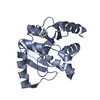
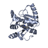
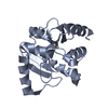
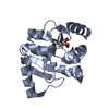
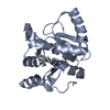
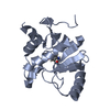
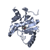
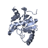
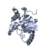
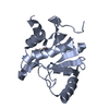
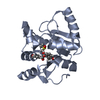
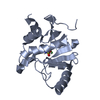
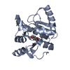
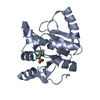
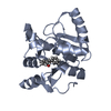
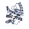
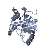
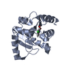
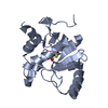
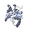
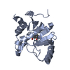
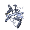
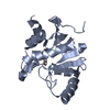
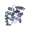
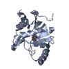
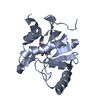
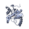
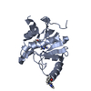
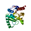
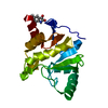
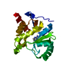
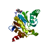
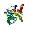
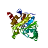
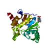
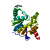
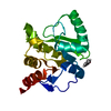
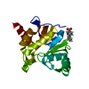
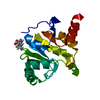
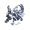
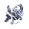
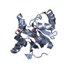
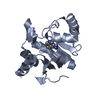
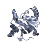
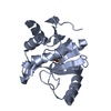
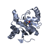
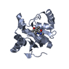
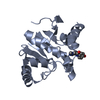
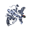
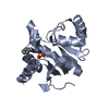
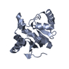
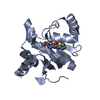
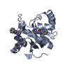
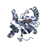
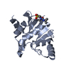
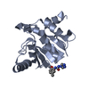
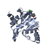
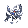
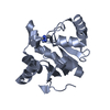
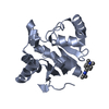
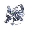
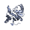
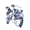
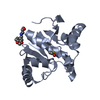
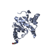
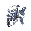
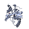
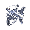
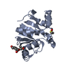
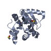
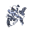
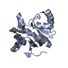
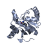
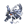
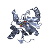
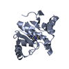
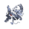
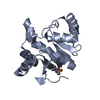
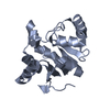
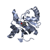
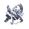
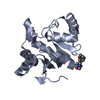
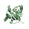
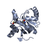
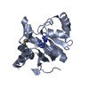
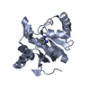
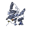
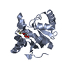
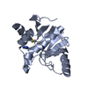
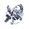
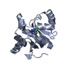
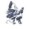
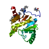
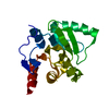
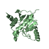
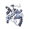
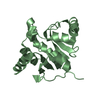
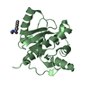
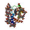
Entry Database : PDB / ID : 7kr1Title Crystal structure of SARS-CoV-2 NSP3 macrodomain (C2 crystal form, 310 K) Non-structural protein 3 Keywords / / / Function / homology Function Domain/homology Component
/ / / / / / / / / / / / / / / / / / / / / / / / / / / / / / / / / / / / / / / / / / / / / / / / / / / / / / / / / / / / / / / / / / / / / / / / / / / / / / / / / / / / / / / / / / / / / / / / / / / / / / / / / / / / / / / / / / / / / / / / / / / / / / / / / / / / / / / / / / / / / / / / / / / / / / / / / / / / / / / / / / / / / / / / / / / / / / / / / / / / Biological species Method / / / Resolution : 1.55 Å Authors Correy, G.J. / Young, I.D. / Thompson, M.C. / Fraser, J.S. Funding support 1items Organization Grant number Country National Science Foundation (NSF, United States) 2031205
Journal : Sci Adv / Year : 2021Title : Fragment binding to the Nsp3 macrodomain of SARS-CoV-2 identified through crystallographic screening and computational docking.Authors: Schuller, M. / Correy, G.J. / Gahbauer, S. / Fearon, D. / Wu, T. / Diaz, R.E. / Young, I.D. / Carvalho Martins, L. / Smith, D.H. / Schulze-Gahmen, U. / Owens, T.W. / Deshpande, I. / Merz, G. ... Authors : Schuller, M. / Correy, G.J. / Gahbauer, S. / Fearon, D. / Wu, T. / Diaz, R.E. / Young, I.D. / Carvalho Martins, L. / Smith, D.H. / Schulze-Gahmen, U. / Owens, T.W. / Deshpande, I. / Merz, G.E. / Thwin, A.C. / Biel, J.T. / Peters, J.K. / Moritz, M. / Herrera, N. / Kratochvil, H.T. / Aimon, A. / Bennett, J.M. / Brandao Neto, J. / Cohen, A.E. / Dias, A. / Douangamath, A. / Dunnett, L. / Fedorov, O. / Ferla, M.P. / Fuchs, M.R. / Gorrie-Stone, T.J. / Holton, J.M. / Johnson, M.G. / Krojer, T. / Meigs, G. / Powell, A.J. / Rack, J.G.M. / Rangel, V.L. / Russi, S. / Skyner, R.E. / Smith, C.A. / Soares, A.S. / Wierman, J.L. / Zhu, K. / O'Brien, P. / Jura, N. / Ashworth, A. / Irwin, J.J. / Thompson, M.C. / Gestwicki, J.E. / von Delft, F. / Shoichet, B.K. / Fraser, J.S. / Ahel, I. History Deposition Nov 18, 2020 Deposition site / Processing site Revision 1.0 Dec 9, 2020 Provider / Type Revision 1.1 Jan 27, 2021 Group / Category / entity_name_comItem / _entity.pdbx_ec / _entity_name_com.nameRevision 1.2 Jun 30, 2021 Group / Category / citation_authorItem _citation.country / _citation.journal_abbrev ... _citation.country / _citation.journal_abbrev / _citation.journal_id_CSD / _citation.journal_id_ISSN / _citation.journal_volume / _citation.pdbx_database_id_DOI / _citation.pdbx_database_id_PubMed / _citation.title / _citation.year Revision 1.3 Oct 18, 2023 Group / Database references / Refinement descriptionCategory chem_comp_atom / chem_comp_bond ... chem_comp_atom / chem_comp_bond / database_2 / pdbx_initial_refinement_model Item / _database_2.pdbx_database_accession
Show all Show less
 Movie
Movie Controller

 Yorodumi
 Open data
 Basic information
 Components
 Keywords
 Function and homology information
 Severe acute respiratory syndrome coronavirus 2
 X-RAY DIFFRACTION /
  Authors
 Citation
 Structure visualization
 Downloads & links
 Other downloads
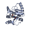


 PDBj
PDBj


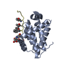
 Assembly

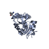
 Mass: 18.015 Da / Num. of mol.: 82 / Source method: isolated from a natural source / Formula: H2O
 Sample preparation
 / Beamline: 8.3.1 / Wavelength: 1.03314 Å
 Processing