 Movie
Movie Controller

[English] 日本語
 Yorodumi
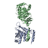
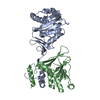
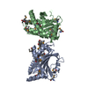
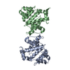
- PDB-7k44: SGBP-B from a complex xyloglucan utilization locus in Bacteroides... -
+ Open data

- Basic information
| Entry | Database: PDB / ID: 7k44 | ||||||
|---|---|---|---|---|---|---|---|
| Title | SGBP-B from a complex xyloglucan utilization locus in Bacteroides uniformis | ||||||
 Components Components | SGBP-B | ||||||
 Keywords Keywords | SUGAR BINDING PROTEIN / surface glycan-binding protein / polysaccharide utilization locus | ||||||
| Function / homology | Surface glycan-binding protein B, xyloglucan binding domain / Surface glycan-binding protein B xyloglucan binding domain / IPT/TIG domain / IPT domain / polysaccharide binding / Immunoglobulin E-set / Immunoglobulin-like fold / metal ion binding / IPT/TIG domain-containing protein Function and homology information Function and homology information | ||||||
| Biological species |  Bacteroides uniformis (bacteria) Bacteroides uniformis (bacteria) | ||||||
| Method |  X-RAY DIFFRACTION / SYNCHROTRON / MOLECULAR REPLACEMENT / Resolution: 1.45 Å X-RAY DIFFRACTION / SYNCHROTRON / MOLECULAR REPLACEMENT / Resolution: 1.45 Å | ||||||
 Authors Authors | Brumer, H. / Van Petegem, F. / Grondin, J.M. | ||||||
| Funding support |  Canada, 1items Canada, 1items
| ||||||
 Citation Citation | Journal: Appl.Environ.Microbiol. / Year: 2022 Title: Cell Surface Xyloglucan Recognition and Hydrolysis by the Human Gut Commensal Bacteroides uniformis. Authors: Grondin, J.M. / Dejean, G. / Van Petegem, F. / Brumer, H. | ||||||
| History |
|
- Structure visualization
| Structure viewer | Molecule: MolmilJmol/JSmol |
|---|
- Downloads & links
-Download
| PDBx/mmCIF format | 7k44.cif.gz | 113.8 KB | Display | PDBx/mmCIF format |
|---|---|---|---|---|
| PDB format | pdb7k44.ent.gz | 68.1 KB | Display | PDB format |
| PDBx/mmJSON format | 7k44.json.gz | Tree view | PDBx/mmJSON format | |
| Others |  Other downloads Other downloads |
-Validation report
| Arichive directory | https://data.pdbj.org/pub/pdb/validation_reports/k4/7k44ftp://data.pdbj.org/pub/pdb/validation_reports/k4/7k44 | HTTPS FTP |
|---|
-Related structure data
| Related structure data |  5e7gS S: Starting model for refinement |
|---|---|
| Similar structure data |


-Links
 PDBj
PDBj
- Assembly
| Deposited unit | 
| ||||||||||||
|---|---|---|---|---|---|---|---|---|---|---|---|---|---|
| 1 |
| ||||||||||||
| Unit cell |
| ||||||||||||
| Components on special symmetry positions |
|
-Components
| #1: Protein | Mass: 42614.391 Da / Num. of mol.: 1 Source method: isolated from a genetically manipulated source Source: (gene. exp.) Bacteroides uniformis (strain ATCC 8492 / DSM 6597 / CIP 103695 / JCM 5828 / NCTC 13054 / VPI 0061) (bacteria)Strain: ATCC 8492 / DSM 6597 / CIP 103695 / JCM 5828 / NCTC 13054 / VPI 0061 Gene: BACUNI_03801 / Production host: |
|---|---|
| #2: Chemical | ChemComp-CA /   Mass: 40.078 Da / Num. of mol.: 1 / Source method: obtained synthetically / Formula: Ca Mass: 40.078 Da / Num. of mol.: 1 / Source method: obtained synthetically / Formula: Ca |
| #3: Water | ChemComp-HOH /  Mass: 18.015 Da / Num. of mol.: 368 / Source method: isolated from a natural source / Formula: H2O Mass: 18.015 Da / Num. of mol.: 368 / Source method: isolated from a natural source / Formula: H2O |
| Has ligand of interest | N |
-Experimental details
-Experiment
| Experiment | Method: X-RAY DIFFRACTION / Number of used crystals: 1 |
|---|
- Sample preparation
| Crystal | Density Matthews: 2.46 Å3/Da / Density % sol: 50 % |
|---|---|
| Crystal grow | Temperature: 298 K / Method: vapor diffusion, sitting drop Details: 0.2M magnesium chloride, 0.1M Tris pH 8.5, 25% PEG 3350 |
-Data collection
| Diffraction | Mean temperature: 100 K / Serial crystal experiment: N |
|---|---|
| Diffraction source | Source: SYNCHROTRON / Site: APS  / Beamline: 23-ID-D / Wavelength: 1.0332 Å / Beamline: 23-ID-D / Wavelength: 1.0332 Å |
| Detector | Type: DECTRIS PILATUS3 S 6M / Detector: PIXEL / Date: Nov 30, 2019 |
| Radiation | Monochromator: Si(111) / Protocol: SINGLE WAVELENGTH / Monochromatic (M) / Laue (L): M / Scattering type: x-ray |
| Radiation wavelength | Wavelength: 1.0332 Å / Relative weight: 1 |
| Reflection | Resolution: 1.45→34.85 Å / Num. obs: 72049 / % possible obs: 99.17 % / Redundancy: 6.6 % / Biso Wilson estimate: 24.04 Å2 / CC1/2: 0.999 / CC star: 1 / Rmerge(I) obs: 0.05159 / Rpim(I) all: 0.02169 / Rrim(I) all: 0.05606 / Net I/σ(I): 15.92 |
| Reflection shell | Resolution: 1.45→1.506 Å / Redundancy: 5.9 % / Rmerge(I) obs: 1.515 / Mean I/σ(I) obs: 0.92 / Num. unique obs: 41563 / CC1/2: 0.43 / CC star: 0.776 / Rpim(I) all: 0.6697 / Rrim(I) all: 1.66 / % possible all: 97.85 |
- Processing
| Software |
| ||||||||||||||||||||||||||||||||||||||||||||||||||||||||||||||||||||||||||||||||||||||||||||||||||||||||||||||||||||||||||||||||||||||||||||||||||||||||||||||||||||||||||||||||||||||
|---|---|---|---|---|---|---|---|---|---|---|---|---|---|---|---|---|---|---|---|---|---|---|---|---|---|---|---|---|---|---|---|---|---|---|---|---|---|---|---|---|---|---|---|---|---|---|---|---|---|---|---|---|---|---|---|---|---|---|---|---|---|---|---|---|---|---|---|---|---|---|---|---|---|---|---|---|---|---|---|---|---|---|---|---|---|---|---|---|---|---|---|---|---|---|---|---|---|---|---|---|---|---|---|---|---|---|---|---|---|---|---|---|---|---|---|---|---|---|---|---|---|---|---|---|---|---|---|---|---|---|---|---|---|---|---|---|---|---|---|---|---|---|---|---|---|---|---|---|---|---|---|---|---|---|---|---|---|---|---|---|---|---|---|---|---|---|---|---|---|---|---|---|---|---|---|---|---|---|---|---|---|---|---|
| Refinement | Method to determine structure: MOLECULAR REPLACEMENT Starting model: 5E7G Resolution: 1.45→34.85 Å / SU ML: 0.2071 / Cross valid method: FREE R-VALUE / σ(F): 1.35 / Phase error: 24.2294 Stereochemistry target values: GeoStd + Monomer Library + CDL v1.2
| ||||||||||||||||||||||||||||||||||||||||||||||||||||||||||||||||||||||||||||||||||||||||||||||||||||||||||||||||||||||||||||||||||||||||||||||||||||||||||||||||||||||||||||||||||||||
| Solvent computation | Shrinkage radii: 0.9 Å / VDW probe radii: 1.11 Å / Solvent model: FLAT BULK SOLVENT MODEL | ||||||||||||||||||||||||||||||||||||||||||||||||||||||||||||||||||||||||||||||||||||||||||||||||||||||||||||||||||||||||||||||||||||||||||||||||||||||||||||||||||||||||||||||||||||||
| Displacement parameters | Biso mean: 30.45 Å2 | ||||||||||||||||||||||||||||||||||||||||||||||||||||||||||||||||||||||||||||||||||||||||||||||||||||||||||||||||||||||||||||||||||||||||||||||||||||||||||||||||||||||||||||||||||||||
| Refinement step | Cycle: LAST / Resolution: 1.45→34.85 Å
| ||||||||||||||||||||||||||||||||||||||||||||||||||||||||||||||||||||||||||||||||||||||||||||||||||||||||||||||||||||||||||||||||||||||||||||||||||||||||||||||||||||||||||||||||||||||
| Refine LS restraints |
| ||||||||||||||||||||||||||||||||||||||||||||||||||||||||||||||||||||||||||||||||||||||||||||||||||||||||||||||||||||||||||||||||||||||||||||||||||||||||||||||||||||||||||||||||||||||
| LS refinement shell |
|
