 Movie
Movie Controller


+ Open data

- Basic information
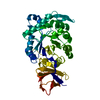
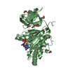
| Entry | Database: PDB / ID: 7jtu | ||||||
|---|---|---|---|---|---|---|---|
| Title | Cytidine deaminase T6S toxin from Pseudomonas syringae | ||||||
 Components Components |
| ||||||
 Keywords Keywords | TOXIN / Immunity / Type VI / T6SS / deaminase | ||||||
| Function / homology |  Function and homology information Function and homology informationImmunity protein Imm5 / Immunity protein Imm5 / : / Single-strand DNA deaminase toxin A, C-terminal / DYW family of nucleic acid deaminases / : / Class I prePAAR effector Rhs1 motif / PAAR motif / PAAR motif Similarity search - Domain/homology | ||||||
| Biological species |  Pseudomonas syringae (bacteria) Pseudomonas syringae (bacteria) | ||||||
| Method |  X-RAY DIFFRACTION / SYNCHROTRON / SAD / Resolution: 3 Å X-RAY DIFFRACTION / SYNCHROTRON / SAD / Resolution: 3 Å | ||||||
 Authors Authors | Bosch, D.E. / Hsu, F. / de Moraes, M.H. / Mougous, J.D. | ||||||
| Funding support |  United States, 1items United States, 1items
| ||||||
 Citation Citation | Journal: Elife / Year: 2021 Title: An interbacterial DNA deaminase toxin directly mutagenizes surviving target populations. Authors: de Moraes, M.H. / Hsu, F. / Huang, D. / Bosch, D.E. / Zeng, J. / Radey, M.C. / Simon, N. / Ledvina, H.E. / Frick, J.P. / Wiggins, P.A. / Peterson, S.B. / Mougous, J.D. | ||||||
| History |
|
- Structure visualization
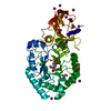
| Structure viewer | Molecule: MolmilJmol/JSmol |
|---|
- Downloads & links
-Download
| PDBx/mmCIF format | 7jtu.cif.gz | 82 KB | Display | PDBx/mmCIF format |
|---|---|---|---|---|
| PDB format | pdb7jtu.ent.gz | 61 KB | Display | PDB format |
| PDBx/mmJSON format | 7jtu.json.gz | Tree view | PDBx/mmJSON format | |
| Others |  Other downloads Other downloads |
-Validation report
| Arichive directory | https://data.pdbj.org/pub/pdb/validation_reports/jt/7jtuftp://data.pdbj.org/pub/pdb/validation_reports/jt/7jtu | HTTPS FTP |
|---|
-Related structure data
| Similar structure data |
|---|


-Links
 PDBj
PDBj- Assembly
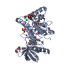
| Deposited unit | 
| ||||||||
|---|---|---|---|---|---|---|---|---|---|
| 1 |
| ||||||||
| Unit cell |
|
-Components
| #1: Protein | Mass: 18559.082 Da / Num. of mol.: 1 Source method: isolated from a genetically manipulated source Source: (gene. exp.) Pseudomonas syringae (bacteria) / Production host: |
|---|---|
| #2: Protein | Mass: 22058.484 Da / Num. of mol.: 1 Source method: isolated from a genetically manipulated source Source: (gene. exp.) Pseudomonas syringae (bacteria) / Production host: |
| #3: Water | ChemComp-HOH /  Mass: 18.015 Da / Num. of mol.: 3 / Source method: isolated from a natural source / Formula: H2O Mass: 18.015 Da / Num. of mol.: 3 / Source method: isolated from a natural source / Formula: H2O |
| Has ligand of interest | N |
| Has protein modification | Y |
-Experimental details
-Experiment
| Experiment | Method: X-RAY DIFFRACTION / Number of used crystals: 1 |
|---|
- Sample preparation
| Crystal | Density Matthews: 5.15 Å3/Da / Density % sol: 76.13 % |
|---|---|
| Crystal grow | Temperature: 293 K / Method: vapor diffusion, hanging drop Details: Hanging drop diffusion. SsdA/SsdAI complex at 10 mg/mL was mixed 1:1 with crystallization solutions containing 20% PEG 3350, 0.1 M Bis-Tris:HCl pH 7.5 and 200 mM MgCl2. Cryopreservation was ...Details: Hanging drop diffusion. SsdA/SsdAI complex at 10 mg/mL was mixed 1:1 with crystallization solutions containing 20% PEG 3350, 0.1 M Bis-Tris:HCl pH 7.5 and 200 mM MgCl2. Cryopreservation was in crystallization solution with 20% glycerol. Temp details: Room temperature |
-Data collection
| Diffraction | Mean temperature: 80 K / Serial crystal experiment: N |
|---|---|
| Diffraction source | Source: SYNCHROTRON / Site: ALS / Beamline: 8.2.2 / Wavelength: 0.979 Å |
| Detector | Type: ADSC QUANTUM 315r / Detector: CCD / Date: Feb 25, 2020 |
| Radiation | Protocol: SINGLE WAVELENGTH / Monochromatic (M) / Laue (L): M / Scattering type: x-ray |
| Radiation wavelength | Wavelength: 0.979 Å / Relative weight: 1 |
| Reflection | Resolution: 3→35 Å / Num. obs: 17597 / % possible obs: 100 % / Redundancy: 28.6 % / Biso Wilson estimate: 79.6 Å2 / Rmerge(I) obs: 0.12 / Net I/σ(I): 22.9 |
| Reflection shell | Resolution: 3→3.03 Å / Redundancy: 29.6 % / Num. unique obs: 434 / Rpim(I) all: 0.279 / % possible all: 100 |
- Processing
| Software |
| |||||||||||||||||||||||||||||||||||||||||||||||||||||||||||||||||||||||||||||||||||||||||||
|---|---|---|---|---|---|---|---|---|---|---|---|---|---|---|---|---|---|---|---|---|---|---|---|---|---|---|---|---|---|---|---|---|---|---|---|---|---|---|---|---|---|---|---|---|---|---|---|---|---|---|---|---|---|---|---|---|---|---|---|---|---|---|---|---|---|---|---|---|---|---|---|---|---|---|---|---|---|---|---|---|---|---|---|---|---|---|---|---|---|---|---|---|
| Refinement | Method to determine structure: SAD / Resolution: 3→33.39 Å / SU ML: 0.39 / Cross valid method: THROUGHOUT / σ(F): 0 / Phase error: 24.99 / Stereochemistry target values: MLHL
| |||||||||||||||||||||||||||||||||||||||||||||||||||||||||||||||||||||||||||||||||||||||||||
| Solvent computation | Shrinkage radii: 0.9 Å / VDW probe radii: 1.11 Å / Solvent model: FLAT BULK SOLVENT MODEL | |||||||||||||||||||||||||||||||||||||||||||||||||||||||||||||||||||||||||||||||||||||||||||
| Displacement parameters | Biso max: 136.64 Å2 / Biso mean: 79.8133 Å2 / Biso min: 42.85 Å2 | |||||||||||||||||||||||||||||||||||||||||||||||||||||||||||||||||||||||||||||||||||||||||||
| Refinement step | Cycle: final / Resolution: 3→33.39 Å
| |||||||||||||||||||||||||||||||||||||||||||||||||||||||||||||||||||||||||||||||||||||||||||
| LS refinement shell | Refine-ID: X-RAY DIFFRACTION / Rfactor Rfree error: 0 / Total num. of bins used: 12
|
