 Movie
Movie Controller

[English] 日本語
 Yorodumi
- PDB-7jrh: X-ray crystal structure of a cyclic peptide containing medin(19-2... -
+ Open data

- Basic information
| Entry | Database: PDB / ID: 7jrh | |||||||||
|---|---|---|---|---|---|---|---|---|---|---|
| Title | X-ray crystal structure of a cyclic peptide containing medin(19-25) and medin(31-37) | |||||||||
 Components Components | Cyclic peptide ASP-GLN-TRP-MLE-GLN-VAL-ASP-ORD-GLU-VAL-THR-GLY-ILE-ILE-THR-ORD | |||||||||
 Keywords Keywords | DE NOVO PROTEIN / medin / cyclic / hairpin / MOF / framework | |||||||||
| Biological species |  Homo sapiens (human) Homo sapiens (human) | |||||||||
| Method |  X-RAY DIFFRACTION / AB INITIO PHASING / Resolution: 1.32 Å X-RAY DIFFRACTION / AB INITIO PHASING / Resolution: 1.32 Å | |||||||||
 Authors Authors | Wierzbicki, M. / Howitz, W.J. / Nowick, J.S. | |||||||||
| Funding support |  United States, 2items United States, 2items
| |||||||||
 Citation Citation | Journal: J.Am.Chem.Soc. / Year: 2020 Title: Interpenetrating Cubes in the X-ray Crystallographic Structure of a Peptide Derived from Medin 19-36 . Authors: Howitz, W.J. / Wierzbicki, M. / Cabanela, R.W. / Saliba, C. / Motavalli, A. / Tran, N. / Nowick, J.S. | |||||||||
| History |
|
- Structure visualization
| Structure viewer | Molecule:  MolmilJmol/JSmol MolmilJmol/JSmol |
|---|
- Downloads & links
-Download
| PDBx/mmCIF format | 7jrh.cif.gz | 21.6 KB | Display | PDBx/mmCIF format |
|---|---|---|---|---|
| PDB format | pdb7jrh.ent.gz | 11.3 KB | Display | PDB format |
| PDBx/mmJSON format | 7jrh.json.gz | Tree view | PDBx/mmJSON format | |
| Others |  Other downloads Other downloads |
-Validation report
| Arichive directory | https://data.pdbj.org/pub/pdb/validation_reports/jr/7jrhftp://data.pdbj.org/pub/pdb/validation_reports/jr/7jrh | HTTPS FTP |
|---|
-Related structure data
| Similar structure data |
|---|
-Links
 PDBj
PDBj
- Assembly
| Deposited unit | 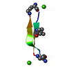
| ||||||||||||||||||
|---|---|---|---|---|---|---|---|---|---|---|---|---|---|---|---|---|---|---|---|
| 1 | x 12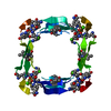
| ||||||||||||||||||
| Unit cell |
| ||||||||||||||||||
| Components on special symmetry positions |
|
-Components
| #1: Protein/peptide | Mass: 1859.084 Da / Num. of mol.: 1 / Source method: obtained synthetically / Source: (synth.) Homo sapiens (human) | ||||||
|---|---|---|---|---|---|---|---|
| #2: Chemical |   Mass: 40.078 Da / Num. of mol.: 2 / Source method: obtained synthetically / Formula: Ca Mass: 40.078 Da / Num. of mol.: 2 / Source method: obtained synthetically / Formula: Ca#3: Water | ChemComp-HOH / |  Mass: 18.015 Da / Num. of mol.: 11 / Source method: isolated from a natural source / Formula: H2O Mass: 18.015 Da / Num. of mol.: 11 / Source method: isolated from a natural source / Formula: H2OHas ligand of interest | N | Has protein modification | Y | |
-Experimental details
-Experiment
| Experiment | Method: X-RAY DIFFRACTION / Number of used crystals: 1 |
|---|
- Sample preparation
| Crystal | Density Matthews: 1.73 Å3/Da / Density % sol: 28.77 % / Description: cube |
|---|---|
| Crystal grow | Temperature: 298 K / Method: vapor diffusion, hanging drop / Details: 0.1 M NaOAc, 0.02M CaCl2, 30% MPD |
-Data collection
| Diffraction | Mean temperature: 100 K / Serial crystal experiment: N |
|---|---|
| Diffraction source | Source: ROTATING ANODE / Type: BRUKER AXS MICROSTAR / Wavelength: 1.54178 Å |
| Detector | Type: APEX II CCD / Detector: CCD / Date: Jul 30, 2020 |
| Radiation | Protocol: SINGLE WAVELENGTH / Monochromatic (M) / Laue (L): M / Scattering type: x-ray |
| Radiation wavelength | Wavelength: 1.54178 Å / Relative weight: 1 |
| Reflection | Resolution: 1.32→30.09 Å / Num. obs: 51865 / % possible obs: 99.74 % / Redundancy: 16.6 % / Biso Wilson estimate: 15.04 Å2 / CC1/2: 1 / CC star: 1 / Rmerge(I) obs: 0.04568 / Rpim(I) all: 0.008085 / Rrim(I) all: 0.04648 / Net I/σ(I): 30.81 |
| Reflection shell | Resolution: 1.321→1.368 Å / Redundancy: 6.4 % / Rmerge(I) obs: 0.3153 / Mean I/σ(I) obs: 3.32 / Num. unique obs: 2021 / CC1/2: 0.925 / CC star: 0.98 / Rpim(I) all: 0.1275 / Rrim(I) all: 0.3423 / % possible all: 97.83 |
- Processing
| Software |
| ||||||||||||||||||||||||||||||||||||||||
|---|---|---|---|---|---|---|---|---|---|---|---|---|---|---|---|---|---|---|---|---|---|---|---|---|---|---|---|---|---|---|---|---|---|---|---|---|---|---|---|---|---|
| Refinement | Method to determine structure: AB INITIO PHASING / Resolution: 1.32→30.04 Å / SU ML: 0.075 / Cross valid method: FREE R-VALUE / σ(F): 1.44 / Phase error: 37.4445 Stereochemistry target values: GeoStd + Monomer Library + CDL v1.2
| ||||||||||||||||||||||||||||||||||||||||
| Solvent computation | Shrinkage radii: 0.9 Å / VDW probe radii: 1.11 Å / Solvent model: FLAT BULK SOLVENT MODEL | ||||||||||||||||||||||||||||||||||||||||
| Displacement parameters | Biso mean: 18.59 Å2 | ||||||||||||||||||||||||||||||||||||||||
| Refinement step | Cycle: LAST / Resolution: 1.32→30.04 Å
| ||||||||||||||||||||||||||||||||||||||||
| Refine LS restraints |
| ||||||||||||||||||||||||||||||||||||||||
| LS refinement shell |
| ||||||||||||||||||||||||||||||||||||||||
| Refinement TLS params. | Method: refined / Origin x: 5.62816389748 Å / Origin y: -5.84557566007 Å / Origin z: -19.9504909138 Å
| ||||||||||||||||||||||||||||||||||||||||
| Refinement TLS group | Selection details: all |
