 Movie
Movie Controller

[English] 日本語
 Yorodumi
- PDB-7jhz: Crystal structure of the carbohydrate-binding domain VP8* of huma... -
+ Open data

- Basic information
| Entry | Database: PDB / ID: 7jhz | ||||||
|---|---|---|---|---|---|---|---|
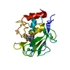
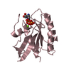
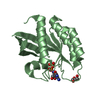
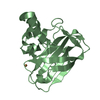
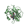
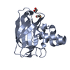
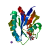
| Title | Crystal structure of the carbohydrate-binding domain VP8* of human P[8] rotavirus strain BM13851 in complex with LNDFH I | ||||||
 Components Components | Outer capsid protein VP4 | ||||||
 Keywords Keywords | VIRAL PROTEIN / Rotavirus / host receptor interaction | ||||||
| Function / homology | Haemagglutinin outer capsid protein VP4, concanavalin-like domain / Outer Capsid protein VP4 (Hemagglutinin) Concanavalin-like domain / viral capsid / Concanavalin A-like lectin/glucanase domain superfamily / symbiont entry into host cell / virion attachment to host cell / Outer capsid protein VP4 Function and homology information Function and homology information | ||||||
| Biological species |  Human rotavirus A Human rotavirus A | ||||||
| Method |  X-RAY DIFFRACTION / SYNCHROTRON / MOLECULAR REPLACEMENT / Resolution: 2.68 Å X-RAY DIFFRACTION / SYNCHROTRON / MOLECULAR REPLACEMENT / Resolution: 2.68 Å | ||||||
 Authors Authors | Xu, S. / Stuckert, M.R. / McGinnis, K.R. / Jiang, X. / Kennedy, M.A. | ||||||
 Citation Citation | Journal: Proc.Natl.Acad.Sci.USA / Year: 2021 Title: Structural basis of P[II] rotavirus evolution and host ranges under selection of histo-blood group antigens. Authors: Xu, S. / McGinnis, K.R. / Liu, Y. / Huang, P. / Tan, M. / Stuckert, M.R. / Burnside, R.E. / Jacob, E.G. / Ni, S. / Jiang, X. / Kennedy, M.A. | ||||||
| History |
|
- Structure visualization
| Structure viewer | Molecule: MolmilJmol/JSmol |
|---|
- Downloads & links
-Download
| PDBx/mmCIF format | 7jhz.cif.gz | 107.9 KB | Display | PDBx/mmCIF format |
|---|---|---|---|---|
| PDB format | pdb7jhz.ent.gz | 81.3 KB | Display | PDB format |
| PDBx/mmJSON format | 7jhz.json.gz | Tree view | PDBx/mmJSON format | |
| Others |  Other downloads Other downloads |
-Validation report
| Arichive directory | https://data.pdbj.org/pub/pdb/validation_reports/jh/7jhzftp://data.pdbj.org/pub/pdb/validation_reports/jh/7jhz | HTTPS FTP |
|---|
-Related structure data
| Related structure data |  6ut9C 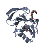 6vkxSC 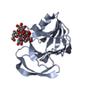 7khuC  7ki5C S: Starting model for refinement C: citing same article ( |
|---|---|
| Similar structure data |


-Links
 PDBj
PDBj- Assembly
| Deposited unit | 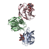
| ||||||||
|---|---|---|---|---|---|---|---|---|---|
| 1 | 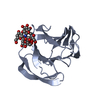
| ||||||||
| 2 | 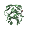
| ||||||||
| 3 | 
| ||||||||
| Unit cell |
|
-Components
| #1: Protein | Mass: 18402.191 Da / Num. of mol.: 3 Source method: isolated from a genetically manipulated source Source: (gene. exp.) Human rotavirus A / Gene: VP8 / Production host:  #2: Polysaccharide | alpha-L-fucopyranose-(1-2)-beta-D-galactopyranose-(1-3)-[alpha-L-fucopyranose-(1-4)]2-acetamido-2- ...alpha-L-fucopyranose-(1-2)-beta-D-galactopyranose-(1-3)-[alpha-L-fucopyranose-(1-4)]2-acetamido-2-deoxy-beta-D-glucopyranose-(1-3)-beta-D-galactopyranose-(1-4)-beta-D-glucopyranose | #3: Polysaccharide | alpha-L-fucopyranose-(1-2)-beta-D-galactopyranose-(1-3)-[alpha-L-fucopyranose-(1-4)]2-acetamido-2- ...alpha-L-fucopyranose-(1-2)-beta-D-galactopyranose-(1-3)-[alpha-L-fucopyranose-(1-4)]2-acetamido-2-deoxy-beta-D-glucopyranose-(1-3)-beta-D-galactopyranose | #4: Chemical |   Mass: 92.094 Da / Num. of mol.: 2 Mass: 92.094 Da / Num. of mol.: 2Source method: isolated from a genetically manipulated source Formula: C3H8O3 #5: Water | ChemComp-HOH / |  Mass: 18.015 Da / Num. of mol.: 9 / Source method: isolated from a natural source / Formula: H2O Mass: 18.015 Da / Num. of mol.: 9 / Source method: isolated from a natural source / Formula: H2OHas ligand of interest | Y | |
|---|
-Experimental details
-Experiment
| Experiment | Method: X-RAY DIFFRACTION / Number of used crystals: 1 |
|---|
- Sample preparation
| Crystal | Density Matthews: 2.33 Å3/Da / Density % sol: 47.33 % |
|---|---|
| Crystal grow | Temperature: 293 K / Method: vapor diffusion, hanging drop / pH: 7.5 Details: 0.1 M HEPES sodium pH 7.5, 2% v/v Polyethylene glycol 400,2.0 M Ammonium sulfate |
-Data collection
| Diffraction | Mean temperature: 100 K / Serial crystal experiment: N | |||||||||||||||||||||
|---|---|---|---|---|---|---|---|---|---|---|---|---|---|---|---|---|---|---|---|---|---|---|
| Diffraction source | Source: SYNCHROTRON / Site: APS  / Beamline: 31-ID / Wavelength: 0.9793 Å / Beamline: 31-ID / Wavelength: 0.9793 Å | |||||||||||||||||||||
| Detector | Type: DECTRIS PILATUS3 S 6M / Detector: PIXEL / Date: Jul 17, 2020 | |||||||||||||||||||||
| Radiation | Protocol: SINGLE WAVELENGTH / Monochromatic (M) / Laue (L): M / Scattering type: x-ray | |||||||||||||||||||||
| Radiation wavelength | Wavelength: 0.9793 Å / Relative weight: 1 | |||||||||||||||||||||
| Reflection | Resolution: 2.68→56.08 Å / Num. obs: 13841 / % possible obs: 95.9 % / Redundancy: 5.1 % / CC1/2: 0.977 / Rmerge(I) obs: 0.183 / Net I/σ(I): 7.1 / Num. measured all: 70641 / Scaling rejects: 164 | |||||||||||||||||||||
| Reflection shell | Diffraction-ID: 1 / Redundancy: 5.3 %
|
- Processing
| Software |
| ||||||||||||||||||||||||||||||||||||||||||||||||||||||||||||
|---|---|---|---|---|---|---|---|---|---|---|---|---|---|---|---|---|---|---|---|---|---|---|---|---|---|---|---|---|---|---|---|---|---|---|---|---|---|---|---|---|---|---|---|---|---|---|---|---|---|---|---|---|---|---|---|---|---|---|---|---|---|
| Refinement | Method to determine structure: MOLECULAR REPLACEMENT Starting model: 6VKX Resolution: 2.68→44.78 Å / Cor.coef. Fo:Fc: 0.903 / Cor.coef. Fo:Fc free: 0.849 / SU B: 12.946 / SU ML: 0.277 / Cross valid method: THROUGHOUT / σ(F): 0 / ESU R Free: 0.389 / Stereochemistry target values: MAXIMUM LIKELIHOOD Details: HYDROGENS HAVE BEEN ADDED IN THE RIDING POSITIONS U VALUES : REFINED INDIVIDUALLY
| ||||||||||||||||||||||||||||||||||||||||||||||||||||||||||||
| Solvent computation | Ion probe radii: 0.8 Å / Shrinkage radii: 0.8 Å / VDW probe radii: 1.2 Å / Solvent model: MASK | ||||||||||||||||||||||||||||||||||||||||||||||||||||||||||||
| Displacement parameters | Biso max: 58.21 Å2 / Biso mean: 23.561 Å2 / Biso min: 5.79 Å2
| ||||||||||||||||||||||||||||||||||||||||||||||||||||||||||||
| Refinement step | Cycle: final / Resolution: 2.68→44.78 Å
| ||||||||||||||||||||||||||||||||||||||||||||||||||||||||||||
| Refine LS restraints |
| ||||||||||||||||||||||||||||||||||||||||||||||||||||||||||||
| LS refinement shell | Resolution: 2.68→2.745 Å / Rfactor Rfree error: 0
|
