 Movie
Movie Controller

[English] 日本語
 Yorodumi
- PDB-7iml: Group deposition for crystallographic fragment screening of SARS-... -
+ Open data

- Basic information
| Entry | Database: PDB / ID: 7iml | ||||||
|---|---|---|---|---|---|---|---|
| Title | Group deposition for crystallographic fragment screening of SARS-CoV-2 nucleocapsid protein (CTD) -- Crystal Structure of SARS-CoV-2 nucleocapsid protein (CTD) in complex with Z1220452176 (Nprot-x0423) | ||||||
 Components Components | Nucleoprotein | ||||||
 Keywords Keywords | VIRAL PROTEIN / Diamond I04-1 / PanDDA2 / XChemExplorer / crystallographic fragment screening / SARS-CoV-2 / nucleocapsid protein / N protein | ||||||
| Function / homology |  Function and homology information Function and homology informationresponse to host immune response / viral RNA genome packaging / negative regulation of interferon-beta production / RNA stem-loop binding / Maturation of nucleoprotein / poly(U) RNA binding / positive regulation of NLRP3 inflammasome complex assembly / MHC class I protein binding / CD28 dependent PI3K/Akt signaling / SARS-CoV-2 targets host intracellular signalling and regulatory pathways ...response to host immune response / viral RNA genome packaging / negative regulation of interferon-beta production / RNA stem-loop binding / Maturation of nucleoprotein / poly(U) RNA binding / positive regulation of NLRP3 inflammasome complex assembly / MHC class I protein binding / CD28 dependent PI3K/Akt signaling / SARS-CoV-2 targets host intracellular signalling and regulatory pathways / VEGFR2 mediated vascular permeability / molecular condensate scaffold activity / MHC class I protein complex / protein sequestering activity / NOD1/2 Signaling Pathway / TAK1-dependent IKK and NF-kappa-B activation / DDX58/IFIH1-mediated induction of interferon-alpha/beta / Interleukin-1 signaling / viral capsid / Interferon alpha/beta signaling / PIP3 activates AKT signaling / viral nucleocapsid / Transcription of SARS-CoV-2 sgRNAs / Translation of Structural Proteins / Virion Assembly and Release / Lectin pathway of complement activation / host cell endoplasmic reticulum-Golgi intermediate compartment / host extracellular region / Induction of Cell-Cell Fusion / Initial triggering of complement / host cell Golgi apparatus / Attachment and Entry / ribonucleoprotein complex / host cell perinuclear region of cytoplasm / symbiont-mediated suppression of host type I interferon-mediated signaling pathway / host cell cytoplasm / SARS-CoV-2 activates/modulates innate and adaptive immune responses / protein homodimerization activity / RNA binding / extracellular region / identical protein binding / cytoplasm Similarity search - Function | ||||||
| Biological species |  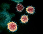 Severe acute respiratory syndrome coronavirus 2SARS-CoV-2 (virus) Severe acute respiratory syndrome coronavirus 2SARS-CoV-2 (virus) | ||||||
| Method |  X-RAY DIFFRACTION / SYNCHROTRON / MOLECULAR REPLACEMENT / Resolution: 1.55 Å X-RAY DIFFRACTION / SYNCHROTRON / MOLECULAR REPLACEMENT / Resolution: 1.55 Å | ||||||
 Authors Authors | Aschenbrenner, J.C. / Luptak, J. / Balcomb, B.H. / Marples, P.G. / Bellini, D. / Yu, C.W. / Douangamath, A. / Dias, A. / Powell, A. / Fearon, D. ...Aschenbrenner, J.C. / Luptak, J. / Balcomb, B.H. / Marples, P.G. / Bellini, D. / Yu, C.W. / Douangamath, A. / Dias, A. / Powell, A. / Fearon, D. / James, L. / von Delft, F. | ||||||
 Citation Citation | Journal: To Be Published Title: Group deposition for crystallographic fragment screening of SARS-CoV-2 nucleocapsid protein (CTD) Authors: Luptak, J. / Aschenbrenner, J.C. / Balcomb, B.H. / Marples, P.G. / Bellini, D. / Yu, C.W. / Douangamath, A. / Dias, A. / Powell, A. / Fearon, D. / James, L. / von Delft, F. | ||||||
| History |
|
- Structure visualization
| Structure viewer | Molecule: MolmilJmol/JSmol |
|---|
- Downloads & links
-Download
| PDBx/mmCIF format | 7iml.cif.gz | 41.5 KB | Display | PDBx/mmCIF format |
|---|---|---|---|---|
| PDB format | pdb7iml.ent.gz | 27.8 KB | Display | PDB format |
| PDBx/mmJSON format | 7iml.json.gz | Tree view | PDBx/mmJSON format | |
| Others |  Other downloads Other downloads |
-Validation report
| Arichive directory | https://data.pdbj.org/pub/pdb/validation_reports/im/7imlftp://data.pdbj.org/pub/pdb/validation_reports/im/7iml | HTTPS FTP |
|---|
-Group deposition
| ID | G_1002353 (51 entries) |
|---|---|
| Title | Group deposition for crystallographic fragment screening of SARS-CoV-2 nucleocapsid protein (CTD) |
| Type | changed state |
| Description | Crystallographic fragment screening of SARS-CoV-2 nucleocapsid protein (CTD) |


-Related structure data
| Related structure data | 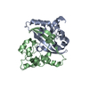 6yunS S: Starting model for refinement |
|---|---|
| Similar structure data |
-Links
 PDBj
PDBj





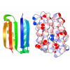
- Assembly
| Deposited unit | 
| ||||||||
|---|---|---|---|---|---|---|---|---|---|
| 1 | 
| ||||||||
| Unit cell |
| ||||||||
| Components on special symmetry positions |
|
-Components
-Protein , 1 types, 1 molecules A
| #1: Protein | Mass: 13118.743 Da / Num. of mol.: 1 Source method: isolated from a genetically manipulated source Source: (gene. exp.) Severe acute respiratory syndrome coronavirus 2Production host:   Escherichia coli (E. coli) / References: UniProt: P0DTC9 Escherichia coli (E. coli) / References: UniProt: P0DTC9 |
|---|
-Non-polymers , 5 types, 168 molecules 

| #2: Chemical | ChemComp-HWH / ~{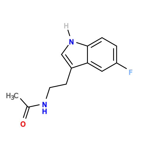 Mass: 220.243 Da / Num. of mol.: 1 Mass: 220.243 Da / Num. of mol.: 1Source method: isolated from a genetically manipulated source Formula: C12H13FN2O / Source: (gene. exp.) SARS-CoV-2 (virus) / Production host: Escherichia coli (E. coli) / Feature type: SUBJECT OF INVESTIGATION |
|---|---|
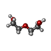
| #3: Chemical | ChemComp-PEG / 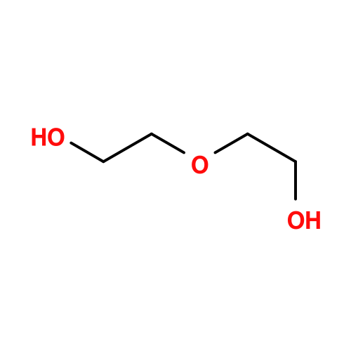 Mass: 106.120 Da / Num. of mol.: 1 Mass: 106.120 Da / Num. of mol.: 1Source method: isolated from a genetically manipulated source Formula: C4H10O3 / Source: (gene. exp.) SARS-CoV-2 (virus) / Production host: Escherichia coli (E. coli) / Feature type: SUBJECT OF INVESTIGATION |
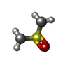
| #4: Chemical | ChemComp-DMS / 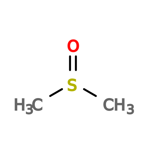 Mass: 78.133 Da / Num. of mol.: 1 Mass: 78.133 Da / Num. of mol.: 1Source method: isolated from a genetically manipulated source Formula: C2H6OS / Source: (gene. exp.) SARS-CoV-2 (virus) / Production host: Escherichia coli (E. coli) / Feature type: SUBJECT OF INVESTIGATION / Comment: DMSO, precipitant*YM |
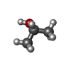
| #5: Chemical | ChemComp-IPA / 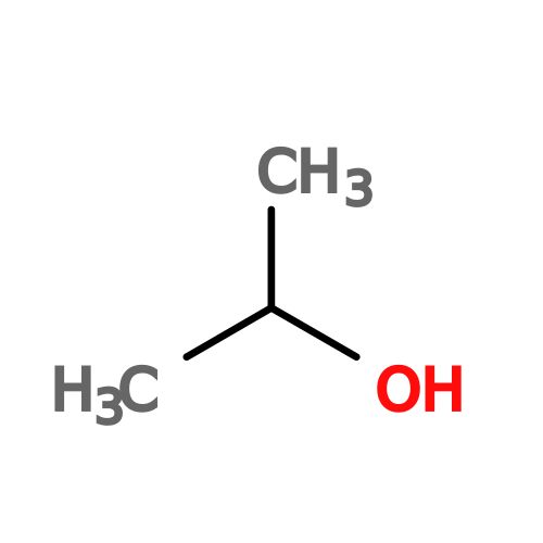 Mass: 60.095 Da / Num. of mol.: 1 / Source method: obtained synthetically / Formula: C3H8O / Feature type: SUBJECT OF INVESTIGATION Mass: 60.095 Da / Num. of mol.: 1 / Source method: obtained synthetically / Formula: C3H8O / Feature type: SUBJECT OF INVESTIGATION |
| #6: Water | ChemComp-HOH / Mass: 18.015 Da / Num. of mol.: 164 / Source method: isolated from a natural source / Formula: H2O |
-Details
| Has ligand of interest | Y |
|---|---|
| Has protein modification | N |
-Experimental details
-Experiment
| Experiment | Method: X-RAY DIFFRACTION / Number of used crystals: 1 |
|---|
- Sample preparation
| Crystal | Density Matthews: 3.03 Å3/Da / Density % sol: 59.47 % |
|---|---|
| Crystal grow | Temperature: 298 K / Method: vapor diffusion, sitting drop / pH: 7.8 Details: 0.1 M HEPES, pH 7.8, 10 % isopropanol, 23 % PEG4000 |
-Data collection
| Diffraction | Mean temperature: 100 K |
|---|---|
| Diffraction source | Source: SYNCHROTRON / Site: Diamond  / Beamline: I04-1 / Wavelength: 0.91261 Å / Beamline: I04-1 / Wavelength: 0.91261 Å |
| Detector | Type: DECTRIS EIGER2 XE 9M / Detector: PIXEL / Date: Aug 12, 2020 |
| Radiation | Protocol: SINGLE WAVELENGTH / Scattering type: x-ray |
| Radiation wavelength | Wavelength: 0.91261 Å / Relative weight: 1 |
| Reflection | Resolution: 1.55→44.138 Å / Num. obs: 11096 / % possible obs: 48.2 % / Redundancy: 6.2 % / Rmerge(I) obs: 0.064 / Rpim(I) all: 0.028 / Rrim(I) all: 0.07 / Net I/σ(I): 12.5 / Num. measured all: 69052 |
| Reflection shell | Resolution: 1.55→1.694 Å / % possible obs: 10.4 % / Redundancy: 4.2 % / Rmerge(I) obs: 0.809 / Num. measured all: 2328 / Num. unique obs: 556 / Rpim(I) all: 0.413 / Rrim(I) all: 0.917 / Net I/σ(I) obs: 1.3 |
- Processing
| Software |
| ||||||||||||||||||||||||||||||||||||||||||||||||||||||||||||||||||||||||||||||||||||||||||||||||||||||||||||||||||||||||||||||||||||||||||||||||||||||||||||||||||||||||||||||||||||||
|---|---|---|---|---|---|---|---|---|---|---|---|---|---|---|---|---|---|---|---|---|---|---|---|---|---|---|---|---|---|---|---|---|---|---|---|---|---|---|---|---|---|---|---|---|---|---|---|---|---|---|---|---|---|---|---|---|---|---|---|---|---|---|---|---|---|---|---|---|---|---|---|---|---|---|---|---|---|---|---|---|---|---|---|---|---|---|---|---|---|---|---|---|---|---|---|---|---|---|---|---|---|---|---|---|---|---|---|---|---|---|---|---|---|---|---|---|---|---|---|---|---|---|---|---|---|---|---|---|---|---|---|---|---|---|---|---|---|---|---|---|---|---|---|---|---|---|---|---|---|---|---|---|---|---|---|---|---|---|---|---|---|---|---|---|---|---|---|---|---|---|---|---|---|---|---|---|---|---|---|---|---|---|---|
| Refinement | Method to determine structure: MOLECULAR REPLACEMENT Starting model: 6YUN Resolution: 1.55→44.18 Å / Cor.coef. Fo:Fc: 0.952 / Cor.coef. Fo:Fc free: 0.918 / SU B: 2.412 / SU ML: 0.087 / Cross valid method: THROUGHOUT / ESU R: 0.234 / ESU R Free: 0.195 / Stereochemistry target values: MAXIMUM LIKELIHOOD / Details: HYDROGENS HAVE BEEN ADDED IN THE RIDING POSITIONS
| ||||||||||||||||||||||||||||||||||||||||||||||||||||||||||||||||||||||||||||||||||||||||||||||||||||||||||||||||||||||||||||||||||||||||||||||||||||||||||||||||||||||||||||||||||||||
| Solvent computation | Ion probe radii: 0.8 Å / Shrinkage radii: 0.8 Å / VDW probe radii: 1.2 Å / Solvent model: MASK | ||||||||||||||||||||||||||||||||||||||||||||||||||||||||||||||||||||||||||||||||||||||||||||||||||||||||||||||||||||||||||||||||||||||||||||||||||||||||||||||||||||||||||||||||||||||
| Displacement parameters | Biso mean: 29.209 Å2
| ||||||||||||||||||||||||||||||||||||||||||||||||||||||||||||||||||||||||||||||||||||||||||||||||||||||||||||||||||||||||||||||||||||||||||||||||||||||||||||||||||||||||||||||||||||||
| Refinement step | Cycle: 1 / Resolution: 1.55→44.18 Å
| ||||||||||||||||||||||||||||||||||||||||||||||||||||||||||||||||||||||||||||||||||||||||||||||||||||||||||||||||||||||||||||||||||||||||||||||||||||||||||||||||||||||||||||||||||||||
| Refine LS restraints |
|