 Movie
Movie Controller

[English] 日本語
 Yorodumi
- PDB-7hjk: PanDDA analysis group deposition -- Crystal structure of ABLE in ... -
+ Open data

- Basic information
| Entry | Database: PDB / ID: 7hjk | ||||||
|---|---|---|---|---|---|---|---|
| Title | PanDDA analysis group deposition -- Crystal structure of ABLE in complex with ZINC000002583439 | ||||||
 Components Components | De novo designed ABLE protein | ||||||
 Keywords Keywords | DE NOVO PROTEIN / Protein design / four-helix bundle / apixaban | ||||||
| Function / homology | (4-fluorophenyl)(pyridin-4-yl)methanone Function and homology information Function and homology information | ||||||
| Biological species | synthetic construct (others) | ||||||
| Method |  X-RAY DIFFRACTION / SYNCHROTRON / MOLECULAR REPLACEMENT / molecular replacement / Resolution: 1.56 Å X-RAY DIFFRACTION / SYNCHROTRON / MOLECULAR REPLACEMENT / molecular replacement / Resolution: 1.56 Å | ||||||
 Authors Authors | Correy, G.J. / Fraser, J.S. | ||||||
| Funding support |  United States, 1items United States, 1items
| ||||||
 Citation Citation | Journal: To be published Title: Fragment screen against ABLE Authors: Correy, G.J. / Fraser, J.S. | ||||||
| History |
|
- Structure visualization
| Structure viewer | Molecule: MolmilJmol/JSmol |
|---|
- Downloads & links
-Download
| PDBx/mmCIF format | 7hjk.cif.gz | 76.7 KB | Display | PDBx/mmCIF format |
|---|---|---|---|---|
| PDB format | pdb7hjk.ent.gz | 61 KB | Display | PDB format |
| PDBx/mmJSON format | 7hjk.json.gz | Tree view | PDBx/mmJSON format | |
| Others |  Other downloads Other downloads |
-Validation report
| Arichive directory | https://data.pdbj.org/pub/pdb/validation_reports/hj/7hjkftp://data.pdbj.org/pub/pdb/validation_reports/hj/7hjk | HTTPS FTP |
|---|
-Group deposition
| ID | G_1002315 (43 entries) |
|---|---|
| Title | PanDDA analysis group deposition of ABLE fragment screen - Enamine Essential fragment library |
| Type | changed state |
| Description | ABLE in complex with fragments identified by X-ray diffraction using ALS 8.3.1, SSRL 12-1 and SSRL 12-2 |


-Related structure data
| Similar structure data |
|---|
-Links
 PDBj
PDBj
- Assembly
| Deposited unit | 
| ||||||||
|---|---|---|---|---|---|---|---|---|---|
| 1 |
| ||||||||
| Unit cell |
|
-Components
| #1: Protein | Mass: 13817.490 Da / Num. of mol.: 1 Source method: isolated from a genetically manipulated source Details: HIS6 purification tag (MHHHHHHENLYFQ) cleaved with TEV protease Source: (gene. exp.) synthetic construct (others) / Plasmid: pET11a / Production host:  | ||||||
|---|---|---|---|---|---|---|---|
| #2: Chemical | 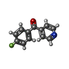  Mass: 201.196 Da / Num. of mol.: 2 / Source method: obtained synthetically / Formula: C12H8FNO Mass: 201.196 Da / Num. of mol.: 2 / Source method: obtained synthetically / Formula: C12H8FNO#3: Water | ChemComp-HOH / |  Mass: 18.015 Da / Num. of mol.: 127 / Source method: isolated from a natural source / Formula: H2O Mass: 18.015 Da / Num. of mol.: 127 / Source method: isolated from a natural source / Formula: H2OHas ligand of interest | Y | Has protein modification | N | |
-Experimental details
-Experiment
| Experiment | Method: X-RAY DIFFRACTION / Number of used crystals: 1 |
|---|
- Sample preparation
| Crystal | Density Matthews: 2.08 Å3/Da / Density % sol: 40.77 % |
|---|---|
| Crystal grow | Temperature: 292 K / Method: vapor diffusion, hanging drop / pH: 5 / Details: 220 mM sodium malonate, 22% PEG 3350 |
-Data collection
| Diffraction | Mean temperature: 100 K | ||||||||||||||||||||||||||||||
|---|---|---|---|---|---|---|---|---|---|---|---|---|---|---|---|---|---|---|---|---|---|---|---|---|---|---|---|---|---|---|---|
| Diffraction source | Source: SYNCHROTRON / Site: ALS / Beamline: 8.3.1 / Wavelength: 1.11583 Å | ||||||||||||||||||||||||||||||
| Detector | Type: DECTRIS PILATUS3 6M / Detector: PIXEL / Date: Mar 23, 2023 | ||||||||||||||||||||||||||||||
| Radiation | Protocol: SINGLE WAVELENGTH / Monochromatic (M) / Laue (L): M / Scattering type: x-ray | ||||||||||||||||||||||||||||||
| Radiation wavelength | Wavelength: 1.11583 Å / Relative weight: 1 | ||||||||||||||||||||||||||||||
| Reflection | Resolution: 1.56→47.11 Å / Num. obs: 15519 / % possible obs: 98.6 % / Redundancy: 3.1 % / Biso Wilson estimate: 18.98 Å2 / CC1/2: 0.999 / Rmerge(I) obs: 0.031 / Rpim(I) all: 0.02 / Rrim(I) all: 0.037 / Net I/σ(I): 18.9 / Num. measured all: 48259 | ||||||||||||||||||||||||||||||
| Reflection shell | Diffraction-ID: 1
|
-Phasing
| Phasing | Method: molecular replacement |
|---|
- Processing
| Software |
| |||||||||||||||||||||||||||||||||||||||||||||||||
|---|---|---|---|---|---|---|---|---|---|---|---|---|---|---|---|---|---|---|---|---|---|---|---|---|---|---|---|---|---|---|---|---|---|---|---|---|---|---|---|---|---|---|---|---|---|---|---|---|---|---|
| Refinement | Method to determine structure: MOLECULAR REPLACEMENT / Resolution: 1.56→45.38 Å / SU ML: 0.14 / Cross valid method: THROUGHOUT / σ(F): 1.34 / Phase error: 25.77 / Stereochemistry target values: ML
| |||||||||||||||||||||||||||||||||||||||||||||||||
| Solvent computation | Shrinkage radii: 0.9 Å / VDW probe radii: 1.1 Å / Solvent model: FLAT BULK SOLVENT MODEL | |||||||||||||||||||||||||||||||||||||||||||||||||
| Displacement parameters | Biso max: 83.32 Å2 / Biso mean: 26.3665 Å2 / Biso min: 10.1 Å2 | |||||||||||||||||||||||||||||||||||||||||||||||||
| Refinement step | Cycle: final / Resolution: 1.56→45.38 Å
| |||||||||||||||||||||||||||||||||||||||||||||||||
| LS refinement shell | Refine-ID: X-RAY DIFFRACTION / Rfactor Rfree error: 0 / Total num. of bins used: 6
|
