 Movie
Movie Controller

[English] 日本語
 Yorodumi
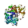
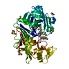
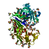
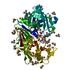
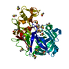
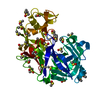
- PDB-7h5q: Crystal structure of endothiapepsin PF_RT2 in complex with TL0015... -
+ Open data

- Basic information
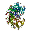
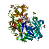
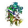
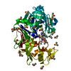
| Entry | Database: PDB / ID: 7h5q | |||||||||
|---|---|---|---|---|---|---|---|---|---|---|
| Title | Crystal structure of endothiapepsin PF_RT2 in complex with TL00150 at 296 K | |||||||||
 Components Components | Endothiapepsin | |||||||||
 Keywords Keywords | HYDROLASE / Endopeptidase / room temperature | |||||||||
| Function / homology |  Function and homology information Function and homology information | |||||||||
| Biological species |  Cryphonectria parasitica (chestnut blight fungus) Cryphonectria parasitica (chestnut blight fungus) | |||||||||
| Method |  X-RAY DIFFRACTION / SYNCHROTRON / MOLECULAR REPLACEMENT / molecular replacement / Resolution: 1.99 Å X-RAY DIFFRACTION / SYNCHROTRON / MOLECULAR REPLACEMENT / molecular replacement / Resolution: 1.99 Å | |||||||||
 Authors Authors | Huang, C.-Y. / Aumonier, S. / Olieric, V. / Wang, M. | |||||||||
| Funding support |  Switzerland, European Union, 2items Switzerland, European Union, 2items
| |||||||||
 Citation Citation | Journal: Acta Crystallogr D Struct Biol / Year: 2024 Title: Cryo2RT: a high-throughput method for room-temperature macromolecular crystallography from cryo-cooled crystals. Authors: Huang, C.Y. / Aumonier, S. / Olieric, V. / Wang, M. | |||||||||
| History |
|
- Structure visualization
| Structure viewer | Molecule: MolmilJmol/JSmol |
|---|
- Downloads & links
-Download
| PDBx/mmCIF format | 7h5q.cif.gz | 78.4 KB | Display | PDBx/mmCIF format |
|---|---|---|---|---|
| PDB format | pdb7h5q.ent.gz | 57.9 KB | Display | PDB format |
| PDBx/mmJSON format | 7h5q.json.gz | Tree view | PDBx/mmJSON format | |
| Others |  Other downloads Other downloads |
-Validation report
| Arichive directory | https://data.pdbj.org/pub/pdb/validation_reports/h5/7h5qftp://data.pdbj.org/pub/pdb/validation_reports/h5/7h5q | HTTPS FTP |
|---|
-Group deposition
| ID | G_1002290 (30 entries) |
|---|---|
| Title | A High-Throughput Method for Room-Temperature Macromolecular Crystallography from Frozen Crystals |
| Type | undefined |
| Description | A High-Throughput Method for Room-Temperature Macromolecular Crystallography from Frozen Crystals |
-Related structure data
| Similar structure data |
|---|
-Links
 PDBj
PDBj
- Assembly
| Deposited unit | 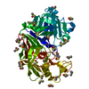
| ||||||||
|---|---|---|---|---|---|---|---|---|---|
| 1 |
| ||||||||
| Unit cell |
|
-Components
| #1: Protein | Mass: 33813.855 Da / Num. of mol.: 1 / Source method: isolated from a natural source Source: (natural) Cryphonectria parasitica (chestnut blight fungus)References: UniProt: P11838, endothiapepsin | ||||||
|---|---|---|---|---|---|---|---|
| #2: Chemical | ChemComp-ACT /   Mass: 59.044 Da / Num. of mol.: 1 / Source method: obtained synthetically / Formula: C2H3O2 Mass: 59.044 Da / Num. of mol.: 1 / Source method: obtained synthetically / Formula: C2H3O2 | ||||||
| #3: Chemical | ChemComp-DMS /   Mass: 78.133 Da / Num. of mol.: 36 / Source method: obtained synthetically / Formula: C2H6OS / Comment: DMSO, precipitant*YM Mass: 78.133 Da / Num. of mol.: 36 / Source method: obtained synthetically / Formula: C2H6OS / Comment: DMSO, precipitant*YM#4: Water | ChemComp-HOH / |  Mass: 18.015 Da / Num. of mol.: 141 / Source method: isolated from a natural source / Formula: H2O Mass: 18.015 Da / Num. of mol.: 141 / Source method: isolated from a natural source / Formula: H2OHas ligand of interest | Y | Has protein modification | Y | |
-Experimental details
-Experiment
| Experiment | Method: X-RAY DIFFRACTION / Number of used crystals: 1 |
|---|
- Sample preparation
| Crystal | Density Matthews: 2.53 Å3/Da / Density % sol: 51.37 % |
|---|---|
| Crystal grow | Temperature: 293 K / Method: vapor diffusion, sitting drop / pH: 4.6 Details: 0.1 M ammonium acetate, 0.1 M sodium acetate pH 4.6, and 26 - 30% (v/v) PEG 4000 |
-Data collection
| Diffraction | Mean temperature: 296 K |
|---|---|
| Diffraction source | Source: SYNCHROTRON / Site: SLS / Beamline: X10SA / Wavelength: 1 Å |
| Detector | Type: DECTRIS EIGER X 16M / Detector: PIXEL / Date: Sep 15, 2023 |
| Radiation | Protocol: SINGLE WAVELENGTH / Monochromatic (M) / Laue (L): M / Scattering type: x-ray |
| Radiation wavelength | Wavelength: 1 Å / Relative weight: 1 |
| Reflection | Resolution: 1.99→43.3 Å / Num. obs: 23124 / % possible obs: 99.7 % / Redundancy: 6.96 % / Biso Wilson estimate: 38.86 Å2 / Rmerge F all: 0.19 / CC1/2: 0.99 / Net I/σ(I): 7.35 |
| Reflection shell | Resolution: 1.99→2.08 Å / Redundancy: 7.07 % / Rmerge F all: 1.32 / Num. unique obs: 3685 / CC1/2: 0.62 / Net I/σ(I) all: 1.52 / % possible all: 99.6 |
-Phasing
| Phasing | Method: molecular replacement |
|---|
- Processing
| Software |
| ||||||||||||||||||||||||||||||||||||||||||||||||||||||||||||||||||||||||||||||||||||||||||||||||||||||||||||
|---|---|---|---|---|---|---|---|---|---|---|---|---|---|---|---|---|---|---|---|---|---|---|---|---|---|---|---|---|---|---|---|---|---|---|---|---|---|---|---|---|---|---|---|---|---|---|---|---|---|---|---|---|---|---|---|---|---|---|---|---|---|---|---|---|---|---|---|---|---|---|---|---|---|---|---|---|---|---|---|---|---|---|---|---|---|---|---|---|---|---|---|---|---|---|---|---|---|---|---|---|---|---|---|---|---|---|---|---|---|
| Refinement | Method to determine structure: MOLECULAR REPLACEMENT / Resolution: 1.99→43.3 Å / Cor.coef. Fo:Fc: 0.958 / Cor.coef. Fo:Fc free: 0.939 / SU R Cruickshank DPI: 0.163 / Cross valid method: THROUGHOUT / σ(F): 0 / SU R Blow DPI: 0.167 / SU Rfree Blow DPI: 0.147 / SU Rfree Cruickshank DPI: 0.146
| ||||||||||||||||||||||||||||||||||||||||||||||||||||||||||||||||||||||||||||||||||||||||||||||||||||||||||||
| Displacement parameters | Biso max: 151.8 Å2 / Biso mean: 42.13 Å2 / Biso min: 26.1 Å2
| ||||||||||||||||||||||||||||||||||||||||||||||||||||||||||||||||||||||||||||||||||||||||||||||||||||||||||||
| Refine analyze | Luzzati coordinate error obs: 0.23 Å | ||||||||||||||||||||||||||||||||||||||||||||||||||||||||||||||||||||||||||||||||||||||||||||||||||||||||||||
| Refinement step | Cycle: final / Resolution: 1.99→43.3 Å
| ||||||||||||||||||||||||||||||||||||||||||||||||||||||||||||||||||||||||||||||||||||||||||||||||||||||||||||
| Refine LS restraints |
| ||||||||||||||||||||||||||||||||||||||||||||||||||||||||||||||||||||||||||||||||||||||||||||||||||||||||||||
| LS refinement shell | Resolution: 1.99→2 Å / Rfactor Rfree error: 0 / Total num. of bins used: 51
|
