[日本語] English
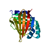
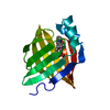
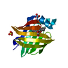
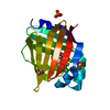
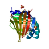
- PDB-7fzx: Crystal Structure of human FABP4 in complex with (2S)-6-(4-chloro... -

+
データを開く


IDまたはキーワード:

読み込み中...

-
基本情報

登録情報
データベース: PDB / ID: 7fzx
タイトルCrystal Structure of human FABP4 in complex with (2S)-6-(4-chlorophenoxy)-2-(4-methylphenoxy)hexanoic acid, i.e. SMILES c1(O[C@@H](CCCCOc2ccc(cc2)Cl)C(=O)O)ccc(cc1)C with IC50=0.585 microM
要素Fatty acid-binding protein, adipocyte
キーワードLIPID BINDING PROTEIN / FATTY ACID BINDING PROTEIN / CYTOPLASM / LIPID-BINDING / TRANSPORT / PROTEIN BINDING
機能・相同性
機能・相同性情報


hormone receptor binding / long-chain fatty acid transmembrane transporter activity / long-chain fatty acid binding / cellular response to lithium ion / Triglyceride catabolism / white fat cell differentiation / long-chain fatty acid transport / fatty acid transport / brown fat cell differentiation / lipid droplet ...hormone receptor binding / long-chain fatty acid transmembrane transporter activity / long-chain fatty acid binding / cellular response to lithium ion / Triglyceride catabolism / white fat cell differentiation / long-chain fatty acid transport / fatty acid transport / brown fat cell differentiation / lipid droplet / cholesterol homeostasis / fatty acid binding / response to bacterium / Transcriptional regulation of white adipocyte differentiation / positive regulation of inflammatory response / cellular response to tumor necrosis factor / positive regulation of cold-induced thermogenesis / MLL4 and MLL3 complexes regulate expression of PPARG target genes in adipogenesis and hepatic steatosis / negative regulation of DNA-templated transcription / extracellular exosome / nucleus / cytosol / cytoplasm
類似検索 - 分子機能
Cytosolic fatty-acid binding proteins signature. / Intracellular lipid binding protein / Cytosolic fatty-acid binding / Lipocalin / cytosolic fatty-acid binding protein family / Lipocalin/cytosolic fatty-acid binding domain / Calycin beta-barrel core domain / Calycin / Lipocalin / Beta Barrel / Mainly Beta
類似検索 - ドメイン・相同性
FORMIC ACID / Chem-WIC / Fatty acid-binding protein, adipocyte
類似検索 - 構成要素
生物種Homo sapiens (ヒト)
手法X線回折 / シンクロトロン / 分子置換 / 解像度: 1.07 Å
データ登録者Ehler, A. / Benz, J. / Obst, U. / Rudolph, M.G.
資金援助 スイス, 1件
組織認可番号
F. Hoffmann-La Roche LTD スイス
引用ジャーナル: Acta Crystallogr D Struct Biol / : 2025
タイトル: A high-resolution data set of fatty acid-binding protein structures. III. Unexpectedly high occurrence of wrong ligands.
著者: Ehler, A. / Bartelmus, C. / Benz, J. / Plitzko, I. / Rudolph, M.G.
履歴
登録2023年4月27日登録サイト: RCSB / 処理サイト: RCSB
改定 1.02023年6月14日Provider: repository / タイプ: Initial release
改定 1.12024年4月3日Group: Data collection / Refinement description
カテゴリ: chem_comp_atom / chem_comp_bond / pdbx_initial_refinement_model
改定 1.22024年11月20日Group: Structure summary / カテゴリ: pdbx_entry_details / struct
Item: _pdbx_entry_details.has_protein_modification / _struct.title
改定 1.32025年8月13日Group: Database references / カテゴリ: citation / citation_author
Item: _citation.journal_abbrev / _citation.journal_id_CSD ..._citation.journal_abbrev / _citation.journal_id_CSD / _citation.journal_id_ISSN / _citation.journal_volume / _citation.page_first / _citation.page_last / _citation.pdbx_database_id_DOI / _citation.pdbx_database_id_PubMed / _citation.title / _citation.year

-
構造の表示

構造ビューア分子:
MolmilJmol/JSmol

ダウンロードとリンク

-
集合体

登録構造単位
A: Fatty acid-binding protein, adipocyte
ヘテロ分子


分子量 (理論値)分子数
合計 (水以外)15,6095
ポリマ-15,0221
非ポリマー5874
4,179232
1


  • 登録構造と同一
  • 登録者・ソフトウェアが定義した集合体
  • 根拠: gel filtration, elutes as a monomer
タイプ名称対称操作
identity operation1_555x,y,z1
手法PISA
単位格子
Length a, b, c (Å)32.514, 53.749, 75.204
Angle α, β, γ (deg.)90.000, 90.000, 90.000
Int Tables number19
Space group name H-MP212121

-
要素

#1: タンパク質 Fatty acid-binding protein, adipocyte / Adipocyte lipid-binding protein / ALBP / Adipocyte-type fatty acid-binding protein / A-FABP / AFABP ...Adipocyte lipid-binding protein / ALBP / Adipocyte-type fatty acid-binding protein / A-FABP / AFABP / Fatty acid-binding protein 4


分子量: 15022.176 Da / 分子数: 1 / 由来タイプ: 組換発現 / 由来: (組換発現) Homo sapiens (ヒト) / 遺伝子: FABP4 / プラスミド: PET15b / 発現宿主: Escherichia coli BL21(DE3) (大腸菌) / 参照: UniProt: P15090
#2: 化合物 ChemComp-WIC / (2S)-6-(4-chlorophenoxy)-2-(4-methylphenoxy)hexanoic acid


分子量: 348.821 Da / 分子数: 1 / 由来タイプ: 合成 / : C19H21ClO4 / タイプ: SUBJECT OF INVESTIGATION
#3: 化合物 ChemComp-SO4 / SULFATE ION / 硫酸ジアニオン


分子量: 96.063 Da / 分子数: 2 / 由来タイプ: 合成 / : SO4
#4: 化合物 ChemComp-FMT / FORMIC ACID / ギ酸


分子量: 46.025 Da / 分子数: 1 / 由来タイプ: 合成 / : CH2O2
#5: 水 ChemComp-HOH / water


分子量: 18.015 Da / 分子数: 232 / 由来タイプ: 天然 / : H2O
研究の焦点であるリガンドがあるかY
Has protein modificationN

-
実験情報

-
実験

実験手法: X線回折 / 使用した結晶の数: 1

-
試料調製

結晶マシュー密度: 2.19 Å3/Da / 溶媒含有率: 43.76 %
結晶化温度: 293 K / 手法: 蒸気拡散法, シッティングドロップ法 / pH: 7
詳細: protein in 25mM Tris/HCl pH 7.5 100mM NaCl, see also PMID 27658368

-
データ収集

回折平均測定温度: 100 K
放射光源由来: シンクロトロン / サイト: SLS / ビームライン: X10SA / 波長: 0.7 Å
検出器タイプ: PSI PILATUS 6M / 検出器: PIXEL / 日付: 2011年11月28日
放射プロトコル: SINGLE WAVELENGTH / 単色(M)・ラウエ(L): M / 散乱光タイプ: x-ray
放射波長波長: 0.7 Å / 相対比: 1
反射解像度: 1.07→43.73 Å / Num. obs: 59037 / % possible obs: 100 % / 冗長度: 6.49 % / Biso Wilson estimate: 13.809 Å2 / CC1/2: 0.999 / Rmerge(I) obs: 0.063 / Rrim(I) all: 0.073 / Rsym value: 0.063 / Χ2: 0.831 / Net I/σ(I): 11.25 / Num. measured all: 383987 / Scaling rejects: 11
反射 シェル

Diffraction-ID: 1

解像度 (Å)冗長度 (%)Rmerge(I) obsMean I/σ(I) obsNum. measured obsNum. possibleNum. unique obsCC1/2Rrim(I) all% possible all
1.07-1.16.6051.3491.3928396430042990.5551.465100
1.1-1.136.6041.0221.8327894422542240.6841.11100
1.13-1.166.5040.8132.3326437406540650.7810.884100
1.16-1.26.1480.6782.6924346396139600.8360.742100
1.2-1.246.1610.5773.1523688384638450.8790.631100
1.24-1.286.8940.4823.9625796374337420.9290.522100
1.28-1.336.8730.3994.7524730359935980.9430.432100
1.33-1.386.8220.3315.7623802348934890.9550.358100
1.38-1.446.7840.2726.9922508331833180.9660.295100
1.44-1.516.6790.218.5921274318531850.980.228100
1.51-1.66.4350.15111.5619619305130490.990.16499.9
1.6-1.695.8040.11713.8116790289328930.9920.129100
1.69-1.816.4680.08619.1617747274427440.9960.094100
1.81-1.956.8060.06226.5317219253025300.9980.067100
1.95-2.146.6890.04834.0315660234123410.9980.052100
2.14-2.396.4790.0439.113859213921390.9990.043100
2.39-2.766.20.03442.9811842191319100.9990.03799.8
2.76-3.385.4880.02747.798934163116280.9990.0399.8
3.38-4.796.6730.02360.518601128912890.9990.025100
4.79-43.736.2040.02357.7548457837810.9990.02699.7

-
解析

ソフトウェア
名称バージョン分類NB
XSCALEデータスケーリング
REFMAC5.6.0119精密化
PDB_EXTRACT3.27データ抽出
XDSデータ削減
PHASER位相決定
精密化構造決定の手法: 分子置換
開始モデル: inhouse model

解像度: 1.07→43.73 Å / Cor.coef. Fo:Fc: 0.979 / Cor.coef. Fo:Fc free: 0.968 / SU B: 1.05 / SU ML: 0.023 / 交差検証法: THROUGHOUT / σ(F): 0 / ESU R: 0.03 / ESU R Free: 0.032 / 立体化学のターゲット値: MAXIMUM LIKELIHOOD
詳細: was registered as 2013380 - structure defined by electron density and confirmed by NMR two conformations with different placement of chloro-phenoxy group
Rfactor反射数%反射Selection details
Rfree0.1779 2867 5 %RANDOM
Rwork0.1472 ---
obs0.1488 54028 96.48 %-
溶媒の処理イオンプローブ半径: 0.8 Å / 減衰半径: 0.8 Å / VDWプローブ半径: 1.2 Å / 溶媒モデル: MASK
原子変位パラメータBiso max: 53.99 Å2 / Biso mean: 14.686 Å2 / Biso min: 6.49 Å2
Baniso -1Baniso -2Baniso -3
1-0.48 Å20 Å20 Å2
2---0.2 Å20 Å2
3----0.28 Å2
精密化ステップサイクル: final / 解像度: 1.07→43.73 Å
タンパク質核酸リガンド溶媒全体
原子数1040 0 101 233 1374
Biso mean--17.74 26.81 -
残基数----133
拘束条件
Refine-IDタイプDev idealDev ideal target
X-RAY DIFFRACTIONr_bond_refined_d0.0240.021212
X-RAY DIFFRACTIONr_bond_other_d0.0020.021218
X-RAY DIFFRACTIONr_angle_refined_deg2.2442.0091644
X-RAY DIFFRACTIONr_angle_other_deg0.8983.0012787
X-RAY DIFFRACTIONr_dihedral_angle_1_deg6.4675160
X-RAY DIFFRACTIONr_dihedral_angle_2_deg31.32725.10249
X-RAY DIFFRACTIONr_dihedral_angle_3_deg12.85815237
X-RAY DIFFRACTIONr_dihedral_angle_4_deg16.574156
X-RAY DIFFRACTIONr_chiral_restr0.1390.2186
X-RAY DIFFRACTIONr_gen_planes_refined0.0110.021336
X-RAY DIFFRACTIONr_gen_planes_other0.0010.02257
X-RAY DIFFRACTIONr_rigid_bond_restr6.33832430
X-RAY DIFFRACTIONr_sphericity_free26.07545
X-RAY DIFFRACTIONr_sphericity_bonded10.48652588
LS精密化 シェル解像度: 1.07→1.098 Å / Rfactor Rfree error: 0 / Total num. of bins used: 20
Rfactor反射数%反射
Rfree0.32 179 -
Rwork0.316 3522 -
all-3701 -
obs--90.44 %

+
万見について

-
お知らせ

-
2022年2月9日: EMDBエントリの付随情報ファイルのフォーマットが新しくなりました

EMDBエントリの付随情報ファイルのフォーマットが新しくなりました

  • EMDBのヘッダファイルのバージョン3が、公式のフォーマットとなりました。
  • これまでは公式だったバージョン1.9は、アーカイブから削除されます。

関連情報:EMDBヘッダ

外部リンク:wwPDBはEMDBデータモデルのバージョン3へ移行します

-
2020年8月12日: 新型コロナ情報

新型コロナ情報

URL: https://pdbj.org/emnavi/covid19.php

新ページ: EM Navigatorに新型コロナウイルスの特設ページを開設しました。

関連情報:Covid-19情報 / 2020年3月5日: 新型コロナウイルスの構造データ

+
2020年3月5日: 新型コロナウイルスの構造データ

新型コロナウイルスの構造データ

関連情報:万見生物種 / 2020年8月12日: 新型コロナ情報

外部リンク:COVID-19特集ページ - PDBj / 今月の分子2020年2月:コロナウイルスプロテーアーゼ

+
2019年1月31日: EMDBのIDの桁数の変更

EMDBのIDの桁数の変更

  • EMDBエントリに付与されているアクセスコード(EMDB-ID)は4桁の数字(例、EMD-1234)でしたが、間もなく枯渇します。これまでの4桁のID番号は4桁のまま変更されませんが、4桁の数字を使い切った後に発行されるIDは5桁以上の数字(例、EMD-12345)になります。5桁のIDは2019年の春頃から発行される見通しです。
  • EM Navigator/万見では、接頭語「EMD-」は省略されています。

関連情報:Q: 「EMD」とは何ですか? / 万見/EM NavigatorにおけるID/アクセスコードの表記

外部リンク:EMDB Accession Codes are Changing Soon! / PDBjへお問い合わせ

+
2017年7月12日: PDB大規模アップデート

PDB大規模アップデート

  • 新バージョンのPDBx/mmCIF辞書形式に基づくデータがリリースされました。
  • 今回の更新はバージョン番号が4から5になる大規模なもので、全エントリデータの書き換えが行われる「Remediation」というアップデートに該当します。
  • このバージョンアップで、電子顕微鏡の実験手法に関する多くの項目の書式が改定されました(例:em_softwareなど)。
  • EM NavigatorとYorodumiでも、この改定に基づいた表示内容になります。

外部リンク:wwPDB Remediation / OneDepデータ基準に準拠した、より強化された内容のモデル構造ファイルが、PDBアーカイブで公開されました。

-
万見 (Yorodumi)

幾万の構造データを、幾万の視点から

  • 万見(Yorodumi)は、EMDB/PDB/SASBDBなどの構造データを閲覧するためのページです。
  • EM Navigatorの詳細ページの後継、Omokage検索のフロントエンドも兼ねています。

関連情報:EMDB / PDB / SASBDB / 3つのデータバンクの比較 / 万見検索 / 2016年8月31日: 新しいEM Navigatorと万見 / 万見文献 / Jmol/JSmol / 機能・相同性情報 / 新しいEM Navigatorと万見の変更点

他の情報も見る