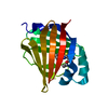
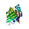
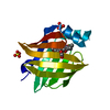
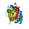
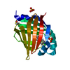
- PDB-7fxd: Crystal Structure of human FABP5 in complex with 2-(indole-1-carb... -
+
データを開く
IDまたはキーワード:
読み込み中...
-
基本情報
登録情報
データベース: PDB / ID: 7fxd
タイトル
Crystal Structure of human FABP5 in complex with 2-(indole-1-carbonylamino)benzoic acid, i.e. SMILES c12N(C(=O)Nc3c(cccc3)C(=O)O)C=Cc1cccc2 with IC50=18.1696 microM
要素
Fatty acid-binding protein 5
キーワード
LIPID BINDING PROTEIN / FATTY ACID BINDING PROTEIN / CYTOPLASM / LIPID-BINDING / TRANSPORT / PROTEIN BINDING
機能・相同性
機能・相同性情報
regulation of prostaglandin biosynthetic process / regulation of retrograde trans-synaptic signaling by endocanabinoid / lipid transport across blood-brain barrier / retrograde trans-synaptic signaling by endocannabinoid / positive regulation of peroxisome proliferator activated receptor signaling pathway / negative regulation of D-glucose transmembrane transport / regulation of sensory perception of pain / phosphatidylcholine biosynthetic process / retinoic acid binding / Differentiation of Keratinocytes in Interfollicular Epidermis in Mammalian Skin ...regulation of prostaglandin biosynthetic process / regulation of retrograde trans-synaptic signaling by endocanabinoid / lipid transport across blood-brain barrier / retrograde trans-synaptic signaling by endocannabinoid / positive regulation of peroxisome proliferator activated receptor signaling pathway / negative regulation of D-glucose transmembrane transport / regulation of sensory perception of pain / phosphatidylcholine biosynthetic process / retinoic acid binding / Differentiation of Keratinocytes in Interfollicular Epidermis in Mammalian Skin / Signaling by Retinoic Acid / long-chain fatty acid transmembrane transporter activity / Triglyceride catabolism / epidermis development / long-chain fatty acid transport / postsynaptic cytosol / fatty acid transport / postsynaptic density, intracellular component / secretory granule membrane / fatty acid binding / lipid metabolic process / glucose metabolic process / azurophil granule lumen / glucose homeostasis / positive regulation of cold-induced thermogenesis / postsynaptic density / lipid binding / synapse / Neutrophil degranulation / glutamatergic synapse / extracellular exosome / extracellular region / nucleoplasm / identical protein binding / nucleus / plasma membrane / cytosol / cytoplasm 類似検索 - 分子機能
構造決定の手法: 分子置換 開始モデル: inhouse model 解像度: 2.44→46.57 Å / Cor.coef. Fo:Fc: 0.9374 / Cor.coef. Fo:Fc free: 0.9224 / SU R Cruickshank DPI: 0.411 / 交差検証法: THROUGHOUT / σ(F): 0 / SU R Blow DPI: 0.48 / SU Rfree Blow DPI: 0.291 / SU Rfree Cruickshank DPI: 0.282 詳細: unidentified electron density close to ligand - cannot be modeled by water
 ムービー
ムービー コントローラー


 データを開く
 基本情報
基本情報 要素
要素 キーワード
キーワード 機能・相同性情報
 Homo sapiens (ヒト)
 X線回折 /
  データ登録者
 スイス, 1件
  引用
引用 構造の表示
構造の表示 ダウンロードとリンク
 その他のダウンロード


 PDBj
PDBj


 集合体
集合体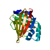


 分子量: 282.294 Da / 分子数: 1 / 由来タイプ: 合成 / 式: C16H14N2O3 / タイプ: SUBJECT OF INVESTIGATION
 分子量: 18.015 Da / 分子数: 19 / 由来タイプ: 天然 / 式: H2O
 試料調製
試料調製 解析
解析