 Movie
Movie Controller

[English] 日本語
 Yorodumi
- PDB-7fjg: Crystal structure of butanol dehydrogenase A (YqdH) in complex wi... -
+ Open data

- Basic information
| Entry | Database: PDB / ID: 7fjg | ||||||
|---|---|---|---|---|---|---|---|
| Title | Crystal structure of butanol dehydrogenase A (YqdH) in complex with partial NADH from Fusobacterium nucleatum | ||||||
 Components Components | NADH-dependent butanol dehydrogenase A | ||||||
 Keywords Keywords | BIOSYNTHETIC PROTEIN / YqdH / partial NADH | ||||||
| Function / homology |  Function and homology information Function and homology informationbutanol dehydrogenase (NAD+) activity / methylglyoxal reductase (NADPH) (acetol producing) activity / alcohol dehydrogenase (NADP+) activity / Oxidoreductases; Acting on the CH-OH group of donors; With NAD+ or NADP+ as acceptor / nucleotide binding / metal ion binding / cytosol Similarity search - Function | ||||||
| Biological species |  Fusobacterium nucleatum subsp. nucleatum (bacteria) Fusobacterium nucleatum subsp. nucleatum (bacteria) | ||||||
| Method |  X-RAY DIFFRACTION / SYNCHROTRON / SAD / Resolution: 2.72 Å X-RAY DIFFRACTION / SYNCHROTRON / SAD / Resolution: 2.72 Å | ||||||
 Authors Authors | Bai, X. / Lan, J. / Wang, L. / Bu, T. / Xu, Y. | ||||||
| Funding support |  Korea, Republic Of, 1items Korea, Republic Of, 1items
| ||||||
 Citation Citation | Journal: To Be Published Title: Crystal structure of butanol dehydrogenase A (YqdH) in complex with partial NADH from Fusobacterium nucleatum Authors: Bai, X. / Lan, J. / Wang, L. / Bu, T. / Xu, Y. | ||||||
| History |
|
- Structure visualization
| Structure viewer | Molecule: MolmilJmol/JSmol |
|---|
- Downloads & links
-Download
| PDBx/mmCIF format | 7fjg.cif.gz | 186.6 KB | Display | PDBx/mmCIF format |
|---|---|---|---|---|
| PDB format | pdb7fjg.ent.gz | 120 KB | Display | PDB format |
| PDBx/mmJSON format | 7fjg.json.gz | Tree view | PDBx/mmJSON format | |
| Others |  Other downloads Other downloads |
-Validation report
| Summary document | 7fjg_validation.pdf.gz | 771.6 KB | Display | wwPDB validaton report |
|---|---|---|---|---|
| Full document | 7fjg_full_validation.pdf.gz | 782 KB | Display | |
| Data in XML | 7fjg_validation.xml.gz | 17 KB | Display | |
| Data in CIF | 7fjg_validation.cif.gz | 22.8 KB | Display | |
| Arichive directory | https://data.pdbj.org/pub/pdb/validation_reports/fj/7fjgftp://data.pdbj.org/pub/pdb/validation_reports/fj/7fjg | HTTPS FTP |
-Related structure data
| Similar structure data |
|---|
-Links
 PDBj
PDBj- Assembly
| Deposited unit | 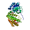
| ||||||||||||
|---|---|---|---|---|---|---|---|---|---|---|---|---|---|
| 1 | 
| ||||||||||||
| Unit cell |
|
-Components
| #1: Protein | Mass: 43336.102 Da / Num. of mol.: 1 Source method: isolated from a genetically manipulated source Source: (gene. exp.) Fusobacterium nucleatum subsp. nucleatum (strain ATCC 25586 / DSM 15643 / BCRC 10681 / CIP 101130 / JCM 8532 / KCTC 2640 / LMG 13131 / VPI 4355) (bacteria)Gene: FN1415 / Production host: References: UniProt: Q8R612, Oxidoreductases; Acting on the CH-OH group of donors; With NAD+ or NADP+ as acceptor |
|---|---|
| #2: Chemical | ChemComp-FE /   Mass: 55.845 Da / Num. of mol.: 1 / Source method: obtained synthetically / Formula: Fe / Feature type: SUBJECT OF INVESTIGATION Mass: 55.845 Da / Num. of mol.: 1 / Source method: obtained synthetically / Formula: Fe / Feature type: SUBJECT OF INVESTIGATION |
| #3: Chemical | ChemComp-ADP /   Mass: 427.201 Da / Num. of mol.: 1 / Source method: isolated from a natural source / Formula: C10H15N5O10P2 / Feature type: SUBJECT OF INVESTIGATION / Comment: ADP, energy-carrying molecule*YM Mass: 427.201 Da / Num. of mol.: 1 / Source method: isolated from a natural source / Formula: C10H15N5O10P2 / Feature type: SUBJECT OF INVESTIGATION / Comment: ADP, energy-carrying molecule*YM |
| #4: Water | ChemComp-HOH /  Mass: 18.015 Da / Num. of mol.: 15 / Source method: isolated from a natural source / Formula: H2O Mass: 18.015 Da / Num. of mol.: 15 / Source method: isolated from a natural source / Formula: H2O |
| Has ligand of interest | Y |
-Experimental details
-Experiment
| Experiment | Method: X-RAY DIFFRACTION / Number of used crystals: 1 |
|---|
- Sample preparation
| Crystal | Density Matthews: 3.14 Å3/Da / Density % sol: 60.83 % |
|---|---|
| Crystal grow | Temperature: 279 K / Method: vapor diffusion, hanging drop / pH: 7.5 Details: 1.8M Ammonium phosphate monobasic, 0.1M HEPES pH 7.5 |
-Data collection
| Diffraction | Mean temperature: 100 K / Serial crystal experiment: N |
|---|---|
| Diffraction source | Source: SYNCHROTRON / Site: SSRF  / Beamline: BL17B1 / Wavelength: 0.9796 Å / Beamline: BL17B1 / Wavelength: 0.9796 Å |
| Detector | Type: DECTRIS PILATUS3 X CdTe 2M / Detector: PIXEL / Date: May 26, 2021 |
| Radiation | Protocol: SINGLE WAVELENGTH / Monochromatic (M) / Laue (L): M / Scattering type: x-ray |
| Radiation wavelength | Wavelength: 0.9796 Å / Relative weight: 1 |
| Reflection | Resolution: 2.719→35.53 Å / Num. obs: 14933 / % possible obs: 98.53 % / Redundancy: 12.4 % / Biso Wilson estimate: 53.81 Å2 / Rsym value: 0.233 / Net I/σ(I): 11.875 |
| Reflection shell | Resolution: 2.719→2.817 Å / Num. unique obs: 752 / Rsym value: 1.157 |
- Processing
| Software |
| |||||||||||||||||||||||||||||||||||||||||||||||||||||||||||||||||||||||||||||
|---|---|---|---|---|---|---|---|---|---|---|---|---|---|---|---|---|---|---|---|---|---|---|---|---|---|---|---|---|---|---|---|---|---|---|---|---|---|---|---|---|---|---|---|---|---|---|---|---|---|---|---|---|---|---|---|---|---|---|---|---|---|---|---|---|---|---|---|---|---|---|---|---|---|---|---|---|---|---|
| Refinement | Method to determine structure: SAD / Resolution: 2.72→35.53 Å / SU ML: 0.3656 / Cross valid method: FREE R-VALUE / σ(F): 1.35 / Phase error: 28.4584 Stereochemistry target values: GeoStd + Monomer Library + CDL v1.2
| |||||||||||||||||||||||||||||||||||||||||||||||||||||||||||||||||||||||||||||
| Solvent computation | Shrinkage radii: 0.9 Å / VDW probe radii: 1.11 Å / Solvent model: FLAT BULK SOLVENT MODEL | |||||||||||||||||||||||||||||||||||||||||||||||||||||||||||||||||||||||||||||
| Displacement parameters | Biso mean: 58.07 Å2 | |||||||||||||||||||||||||||||||||||||||||||||||||||||||||||||||||||||||||||||
| Refinement step | Cycle: LAST / Resolution: 2.72→35.53 Å
| |||||||||||||||||||||||||||||||||||||||||||||||||||||||||||||||||||||||||||||
| Refine LS restraints |
| |||||||||||||||||||||||||||||||||||||||||||||||||||||||||||||||||||||||||||||
| LS refinement shell |
|
