 Movie
Movie Controller

[English] 日本語
 Yorodumi
- PDB-7fhm: Crystal structure of an orphan heme uptake protein (MhuP) of ABC ... -
+ Open data

- Basic information
| Entry | Database: PDB / ID: 7fhm | ||||||
|---|---|---|---|---|---|---|---|
| Title | Crystal structure of an orphan heme uptake protein (MhuP) of ABC transporter from Mycobacterium tuberculosis (Form I) | ||||||
 Components Components | Probable periplasmic iron-transport lipoprotein | ||||||
 Keywords Keywords | TRANSPORT PROTEIN / Hemoglobin / Iron homeostasis / Nutritional immunity / substrate-binding protein / nucleotide-binding domain | ||||||
| Function / homology |  Function and homology information Function and homology informationiron coordination entity transport / outer membrane-bounded periplasmic space Similarity search - Function | ||||||
| Biological species |  Mycobacterium tuberculosis H37Rv (bacteria) Mycobacterium tuberculosis H37Rv (bacteria) | ||||||
| Method |  X-RAY DIFFRACTION / MOLECULAR REPLACEMENT / molecular replacement / Resolution: 1.8 Å X-RAY DIFFRACTION / MOLECULAR REPLACEMENT / molecular replacement / Resolution: 1.8 Å | ||||||
 Authors Authors | Mandal, S.K. / Kanaujia, S.P. | ||||||
| Funding support |  India, 1items India, 1items
| ||||||
 Citation Citation | Journal: Int.J.Biol.Macromol. / Year: 2022 Title: Role of an orphan substrate-binding protein MhuP in transient heme transfer in Mycobacterium tuberculosis Authors: Mandal, S.K. / Kanaujia, S.P. | ||||||
| History |
|
- Structure visualization
| Structure viewer | Molecule: MolmilJmol/JSmol |
|---|
- Downloads & links
-Download
| PDBx/mmCIF format | 7fhm.cif.gz | 252.3 KB | Display | PDBx/mmCIF format |
|---|---|---|---|---|
| PDB format | pdb7fhm.ent.gz | 201.2 KB | Display | PDB format |
| PDBx/mmJSON format | 7fhm.json.gz | Tree view | PDBx/mmJSON format | |
| Others |  Other downloads Other downloads |
-Validation report
| Arichive directory | https://data.pdbj.org/pub/pdb/validation_reports/fh/7fhmftp://data.pdbj.org/pub/pdb/validation_reports/fh/7fhm | HTTPS FTP |
|---|
-Related structure data
| Related structure data |  7fhpC  4pm4S S: Starting model for refinement C: citing same article ( |
|---|---|
| Similar structure data |
-Links
 PDBj
PDBj


- Assembly
| Deposited unit | 
| ||||||||||||||||||
|---|---|---|---|---|---|---|---|---|---|---|---|---|---|---|---|---|---|---|---|
| 1 | 
| ||||||||||||||||||
| 2 | 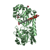
| ||||||||||||||||||
| Unit cell |
| ||||||||||||||||||
| Noncrystallographic symmetry (NCS) | NCS domain:
NCS domain segments: Component-ID: _ / Ens-ID: 1 / Beg auth comp-ID: ALA / Beg label comp-ID: ALA / End auth comp-ID: GLY / End label comp-ID: GLY / Refine code: _ / Auth seq-ID: 9 - 298 / Label seq-ID: 9 - 298
|
-Components
-Protein , 1 types, 2 molecules AB
| #1: Protein | Mass: 32621.182 Da / Num. of mol.: 2 Source method: isolated from a genetically manipulated source Source: (gene. exp.) Mycobacterium tuberculosis H37Rv (bacteria)Strain: H37Rv / Gene: Rv0265c / Plasmid: pET22b / Production host: |
|---|
-Non-polymers , 7 types, 705 molecules 


| #2: Chemical | ChemComp-CIT /  Mass: 192.124 Da / Num. of mol.: 1 / Source method: obtained synthetically / Formula: C6H8O7 Mass: 192.124 Da / Num. of mol.: 1 / Source method: obtained synthetically / Formula: C6H8O7 | ||||||||||
|---|---|---|---|---|---|---|---|---|---|---|---|
| #3: Chemical |  Mass: 62.068 Da / Num. of mol.: 2 / Source method: obtained synthetically / Formula: C2H6O2 Mass: 62.068 Da / Num. of mol.: 2 / Source method: obtained synthetically / Formula: C2H6O2#4: Chemical |  Mass: 92.094 Da / Num. of mol.: 3 / Source method: obtained synthetically / Formula: C3H8O3 Mass: 92.094 Da / Num. of mol.: 3 / Source method: obtained synthetically / Formula: C3H8O3#5: Chemical | ChemComp-CO2 / |  Mass: 44.010 Da / Num. of mol.: 1 / Source method: obtained synthetically / Formula: CO2 Mass: 44.010 Da / Num. of mol.: 1 / Source method: obtained synthetically / Formula: CO2#6: Chemical | ChemComp-CO3 / |  Mass: 60.009 Da / Num. of mol.: 1 / Source method: obtained synthetically / Formula: CO3 Mass: 60.009 Da / Num. of mol.: 1 / Source method: obtained synthetically / Formula: CO3#7: Chemical | ChemComp-ACT / |  Mass: 59.044 Da / Num. of mol.: 1 / Source method: obtained synthetically / Formula: C2H3O2 Mass: 59.044 Da / Num. of mol.: 1 / Source method: obtained synthetically / Formula: C2H3O2#8: Water | ChemComp-HOH / | Mass: 18.015 Da / Num. of mol.: 696 / Source method: isolated from a natural source / Formula: H2O |
-Details
| Has ligand of interest | N |
|---|
-Experimental details
-Experiment
| Experiment | Method: X-RAY DIFFRACTION / Number of used crystals: 1 |
|---|
- Sample preparation
| Crystal | Density Matthews: 2.53 Å3/Da / Density % sol: 51.45 % Description: Monoclinic THE ENTRY CONTAINS FRIEDEL PAIRS IN I/F_PLUS/MINUS COLUMNS. |
|---|---|
| Crystal grow | Temperature: 293 K / Method: microbatch / pH: 5 / Details: 0.1 M citrate pH 5.0 and 20% PEG 6000 |
-Data collection
| Diffraction | Mean temperature: 100 K / Serial crystal experiment: N | ||||||||||||||||||||||||
|---|---|---|---|---|---|---|---|---|---|---|---|---|---|---|---|---|---|---|---|---|---|---|---|---|---|
| Diffraction source | Source: ROTATING ANODE / Type: RIGAKU MICROMAX-007 HF / Wavelength: 1.5418 Å | ||||||||||||||||||||||||
| Detector | Type: RIGAKU RAXIS IV++ / Detector: IMAGE PLATE / Date: Feb 26, 2020 / Details: Vari Max HF | ||||||||||||||||||||||||
| Radiation | Monochromator: Ni / Protocol: SINGLE WAVELENGTH / Monochromatic (M) / Laue (L): M / Scattering type: x-ray | ||||||||||||||||||||||||
| Radiation wavelength | Wavelength: 1.5418 Å / Relative weight: 1 | ||||||||||||||||||||||||
| Reflection | Resolution: 1.8→55.09 Å / Num. obs: 61369 / % possible obs: 100 % / Redundancy: 4.7 % / CC1/2: 0.998 / Rmerge(I) obs: 0.073 / Rpim(I) all: 0.037 / Rrim(I) all: 0.082 / Net I/σ(I): 14.1 | ||||||||||||||||||||||||
| Reflection shell | Diffraction-ID: 1
|
-Phasing
| Phasing | Method: molecular replacement | |||||||||
|---|---|---|---|---|---|---|---|---|---|---|
| Phasing MR | Model details: Phaser MODE: MR_AUTO
|
- Processing
| Software |
| |||||||||||||||||||||||||||||||||||||||||||||||||||||||||||||||||||||||||||
|---|---|---|---|---|---|---|---|---|---|---|---|---|---|---|---|---|---|---|---|---|---|---|---|---|---|---|---|---|---|---|---|---|---|---|---|---|---|---|---|---|---|---|---|---|---|---|---|---|---|---|---|---|---|---|---|---|---|---|---|---|---|---|---|---|---|---|---|---|---|---|---|---|---|---|---|---|
| Refinement | Method to determine structure: MOLECULAR REPLACEMENT Starting model: 4PM4 Resolution: 1.8→55.03 Å / Cor.coef. Fo:Fc: 0.964 / Cor.coef. Fo:Fc free: 0.948 / SU B: 4.256 / SU ML: 0.07 / SU R Cruickshank DPI: 0.107 / Cross valid method: THROUGHOUT / σ(F): 0 / ESU R: 0.107 / ESU R Free: 0.104 / Stereochemistry target values: MAXIMUM LIKELIHOOD Details: U VALUES : WITH TLS ADDED HYDROGENS HAVE BEEN ADDED IN THE RIDING POSITIONS
| |||||||||||||||||||||||||||||||||||||||||||||||||||||||||||||||||||||||||||
| Solvent computation | Ion probe radii: 0.8 Å / Shrinkage radii: 0.8 Å / VDW probe radii: 1.2 Å / Solvent model: MASK | |||||||||||||||||||||||||||||||||||||||||||||||||||||||||||||||||||||||||||
| Displacement parameters | Biso max: 83.47 Å2 / Biso mean: 24.433 Å2 / Biso min: 8.36 Å2
| |||||||||||||||||||||||||||||||||||||||||||||||||||||||||||||||||||||||||||
| Refinement step | Cycle: final / Resolution: 1.8→55.03 Å
| |||||||||||||||||||||||||||||||||||||||||||||||||||||||||||||||||||||||||||
| Refine LS restraints |
| |||||||||||||||||||||||||||||||||||||||||||||||||||||||||||||||||||||||||||
| Refine LS restraints NCS | Ens-ID: 1 / Number: 9218 / Refine-ID: X-RAY DIFFRACTION / Type: interatomic distance / Rms dev position: 0.1 Å / Weight position: 0.05
| |||||||||||||||||||||||||||||||||||||||||||||||||||||||||||||||||||||||||||
| LS refinement shell | Resolution: 1.8→1.847 Å / Rfactor Rfree error: 0 / Total num. of bins used: 20
| |||||||||||||||||||||||||||||||||||||||||||||||||||||||||||||||||||||||||||
| Refinement TLS params. | Method: refined / Refine-ID: X-RAY DIFFRACTION
| |||||||||||||||||||||||||||||||||||||||||||||||||||||||||||||||||||||||||||
| Refinement TLS group |
|
