 Movie
Movie Controller

[English] 日本語
 Yorodumi
- PDB-7fhe: Structure of prenyltransferase mutant Q295F from Streptomyces sp.... -
+ Open data

- Basic information
| Entry | Database: PDB / ID: 7fhe | ||||||
|---|---|---|---|---|---|---|---|
| Title | Structure of prenyltransferase mutant Q295F from Streptomyces sp. (strain CL190) | ||||||
 Components Components | Prenyltransferase | ||||||
 Keywords Keywords | TRANSFERASE / PT-barrel | ||||||
| Function / homology | Aromatic prenyltransferase, CloQ-type / Prenyltransferase-like superfamily / Aromatic prenyltransferase Orf2 / Aromatic prenyltransferase / prenyltransferase activity / metal ion binding / Prenyltransferase Function and homology information Function and homology information | ||||||
| Biological species |  Streptomyces sp. (bacteria) Streptomyces sp. (bacteria) | ||||||
| Method |  X-RAY DIFFRACTION / SYNCHROTRON / MOLECULAR REPLACEMENT / molecular replacement / Resolution: 2.5 Å X-RAY DIFFRACTION / SYNCHROTRON / MOLECULAR REPLACEMENT / molecular replacement / Resolution: 2.5 Å | ||||||
 Authors Authors | Xue, B. / Lim, K.J.H. / Hartono, Y.D. / Go, M.D.K. / Fan, H. / Yew, W.S. | ||||||
| Funding support |  Singapore, 1items Singapore, 1items
| ||||||
 Citation Citation | Journal: Acs Catalysis / Year: 2022 Title: Structure-Guided Engineering of Prenyltransferase NphB for High-Yield and Regioselective Cannabinoid Production. Authors: Lim, K.J.H. / Hartono, Y.D. / Xue, B. / Go, M.K. / Fan, H. / Yew, W.S. | ||||||
| History |
|
- Structure visualization
| Structure viewer | Molecule: MolmilJmol/JSmol |
|---|
- Downloads & links
-Download
| PDBx/mmCIF format | 7fhe.cif.gz | 70.3 KB | Display | PDBx/mmCIF format |
|---|---|---|---|---|
| PDB format | pdb7fhe.ent.gz | 49.2 KB | Display | PDB format |
| PDBx/mmJSON format | 7fhe.json.gz | Tree view | PDBx/mmJSON format | |
| Others |  Other downloads Other downloads |
-Validation report
| Arichive directory | https://data.pdbj.org/pub/pdb/validation_reports/fh/7fheftp://data.pdbj.org/pub/pdb/validation_reports/fh/7fhe | HTTPS FTP |
|---|
-Related structure data
| Related structure data |  7fhbC  7fhcC  7fhdC  7fhfC  1zb6S S: Starting model for refinement C: citing same article ( |
|---|---|
| Similar structure data |
-Links
 PDBj
PDBj- Assembly
| Deposited unit | 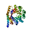
| ||||||||
|---|---|---|---|---|---|---|---|---|---|
| 1 |
| ||||||||
| Unit cell |
|
-Components
| #1: Protein | Mass: 33951.406 Da / Num. of mol.: 1 / Mutation: Q295F Source method: isolated from a genetically manipulated source Source: (gene. exp.) Streptomyces sp. (strain CL190) (bacteria)Strain: CL190 / Plasmid: pSY5CDF / Production host: |
|---|---|
| #2: Water | ChemComp-HOH /  Mass: 18.015 Da / Num. of mol.: 5 / Source method: isolated from a natural source / Formula: H2O Mass: 18.015 Da / Num. of mol.: 5 / Source method: isolated from a natural source / Formula: H2O |
-Experimental details
-Experiment
| Experiment | Method: X-RAY DIFFRACTION / Number of used crystals: 1 |
|---|
- Sample preparation
| Crystal | Density Matthews: 1.95 Å3/Da / Density % sol: 36.91 % / Mosaicity: 0.26 ° |
|---|---|
| Crystal grow | Temperature: 293 K / Method: vapor diffusion, sitting drop / pH: 5 Details: 0.1 M Sodium citrate tribasic dihydrate pH 5.0, 18% w/v PEG 20,000 |
-Data collection
| Diffraction | Mean temperature: 100 K / Serial crystal experiment: N |
|---|---|
| Diffraction source | Source: SYNCHROTRON / Site: Australian Synchrotron  / Beamline: MX1 / Wavelength: 0.95373 Å / Beamline: MX1 / Wavelength: 0.95373 Å |
| Detector | Type: DECTRIS EIGER2 X 9M / Detector: PIXEL / Date: Oct 21, 2020 |
| Radiation | Protocol: SINGLE WAVELENGTH / Monochromatic (M) / Laue (L): M / Scattering type: x-ray |
| Radiation wavelength | Wavelength: 0.95373 Å / Relative weight: 1 |
| Reflection | Resolution: 2.5→29.45 Å / Num. obs: 9690 / % possible obs: 99.9 % / Redundancy: 12.6 % / Biso Wilson estimate: 38.74 Å2 / CC1/2: 0.998 / Rmerge(I) obs: 0.154 / Rpim(I) all: 0.044 / Rrim(I) all: 0.16 / Net I/σ(I): 11.4 / Num. measured all: 122458 / Scaling rejects: 31 |
| Reflection shell | Resolution: 2.5→2.6 Å / Redundancy: 13.1 % / Rmerge(I) obs: 1.015 / Num. unique obs: 1073 / CC1/2: 0.925 / Rpim(I) all: 0.289 / Rrim(I) all: 1.056 / % possible all: 100 |
-Phasing
| Phasing | Method: molecular replacement | |||||||||
|---|---|---|---|---|---|---|---|---|---|---|
| Phasing MR | Model details: Phaser MODE: MR_AUTO
|
- Processing
| Software |
| ||||||||||||||||||||||||
|---|---|---|---|---|---|---|---|---|---|---|---|---|---|---|---|---|---|---|---|---|---|---|---|---|---|
| Refinement | Method to determine structure: MOLECULAR REPLACEMENT Starting model: 1ZB6 Resolution: 2.5→29.45 Å / SU ML: 0.34 / Cross valid method: THROUGHOUT / σ(F): 1.34 / Phase error: 36.79 / Stereochemistry target values: ML
| ||||||||||||||||||||||||
| Solvent computation | Shrinkage radii: 0.9 Å / VDW probe radii: 1.11 Å / Solvent model: FLAT BULK SOLVENT MODEL | ||||||||||||||||||||||||
| Displacement parameters | Biso max: 94.53 Å2 / Biso mean: 42.2289 Å2 / Biso min: 26.86 Å2 | ||||||||||||||||||||||||
| Refinement step | Cycle: final / Resolution: 2.5→29.45 Å
| ||||||||||||||||||||||||
| LS refinement shell | Refine-ID: X-RAY DIFFRACTION / Rfactor Rfree error: 0 / Total num. of bins used: 3 / % reflection obs: 100 %
|
