 Movie
Movie Controller

[English] 日本語
 Yorodumi
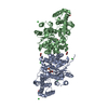
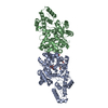
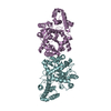
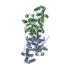
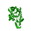
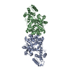
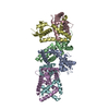
- PDB-7fg6: Crystal structure of the Tyrosyl-tRNA synthetase (TyrRS) in Nanoa... -
+ Open data

- Basic information
| Entry | Database: PDB / ID: 7fg6 | ||||||
|---|---|---|---|---|---|---|---|
| Title | Crystal structure of the Tyrosyl-tRNA synthetase (TyrRS) in Nanoarchaeum equitans | ||||||
 Components Components | Tyrosine--tRNA ligase | ||||||
 Keywords Keywords | LIGASE / TyrRS / tRNA synthetase | ||||||
| Function / homology |  Function and homology information Function and homology informationtyrosyl-tRNA aminoacylation / tyrosine-tRNA ligase / tyrosine-tRNA ligase activity / ATP binding / cytoplasm Similarity search - Function | ||||||
| Biological species |   Nanoarchaeum equitans Kin4-M (archaea) Nanoarchaeum equitans Kin4-M (archaea) | ||||||
| Method |  X-RAY DIFFRACTION / SYNCHROTRON / MOLECULAR REPLACEMENT / Resolution: 2.8 Å X-RAY DIFFRACTION / SYNCHROTRON / MOLECULAR REPLACEMENT / Resolution: 2.8 Å | ||||||
 Authors Authors | Noguchi, H. / Kamata, K. / Park, S.Y. / Tamura, K. | ||||||
| Funding support |  Japan, 1items Japan, 1items
| ||||||
 Citation Citation | Journal: Biochem.Biophys.Res.Commun. / Year: 2021 Title: Crystal structure of Nanoarchaeum equitans tyrosyl-tRNA synthetase and its aminoacylation activity toward tRNA Tyr with an extra guanosine residue at the 5'-terminus. Authors: Horikoshi, T. / Noguchi, H. / Umehara, T. / Mutsuro-Aoki, H. / Kurihara, R. / Noguchi, R. / Hashimoto, T. / Watanabe, Y. / Ando, T. / Kamata, K. / Park, S.Y. / Tamura, K. | ||||||
| History |
|
- Structure visualization
| Structure viewer | Molecule: MolmilJmol/JSmol |
|---|
- Downloads & links
-Download
| PDBx/mmCIF format | 7fg6.cif.gz | 87.6 KB | Display | PDBx/mmCIF format |
|---|---|---|---|---|
| PDB format | pdb7fg6.ent.gz | 63.7 KB | Display | PDB format |
| PDBx/mmJSON format | 7fg6.json.gz | Tree view | PDBx/mmJSON format | |
| Others |  Other downloads Other downloads |
-Validation report
| Summary document | 7fg6_validation.pdf.gz | 428.7 KB | Display | wwPDB validaton report |
|---|---|---|---|---|
| Full document | 7fg6_full_validation.pdf.gz | 434.2 KB | Display | |
| Data in XML | 7fg6_validation.xml.gz | 14.8 KB | Display | |
| Data in CIF | 7fg6_validation.cif.gz | 19.2 KB | Display | |
| Arichive directory | https://data.pdbj.org/pub/pdb/validation_reports/fg/7fg6ftp://data.pdbj.org/pub/pdb/validation_reports/fg/7fg6 | HTTPS FTP |
-Related structure data
| Related structure data | 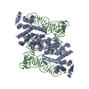 1j1uS S: Starting model for refinement |
|---|---|
| Similar structure data |


-Links
 PDBj
PDBj- Assembly
| Deposited unit | 
| ||||||||||||
|---|---|---|---|---|---|---|---|---|---|---|---|---|---|
| 1 | 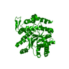
| ||||||||||||
| Unit cell |
|
-Components
| #1: Protein | Mass: 43244.285 Da / Num. of mol.: 1 Source method: isolated from a genetically manipulated source Source: (gene. exp.) Nanoarchaeum equitans Kin4-M (archaea) / Strain: Kin4-M / Gene: tyrS / Production host:  |
|---|---|
| #2: Water | ChemComp-HOH /  Mass: 18.015 Da / Num. of mol.: 11 / Source method: isolated from a natural source / Formula: H2O Mass: 18.015 Da / Num. of mol.: 11 / Source method: isolated from a natural source / Formula: H2O |
-Experimental details
-Experiment
| Experiment | Method: X-RAY DIFFRACTION / Number of used crystals: 1 |
|---|
- Sample preparation
| Crystal | Density Matthews: 2.53 Å3/Da / Density % sol: 51.41 % Description: THE ENTRY CONTAINS FRIEDEL PAIRS IN I/F_PLUS/MINUS COLUMNS. |
|---|---|
| Crystal grow | Temperature: 297.15 K / Method: vapor diffusion, hanging drop / pH: 6.5 Details: 0.2 M Magnesium acetate, 0.1 M Sodium cacodylate pH6.5, 20% (w/v) PEG 8,000 |
-Data collection
| Diffraction | Mean temperature: 93.15 K / Serial crystal experiment: N |
|---|---|
| Diffraction source | Source: SYNCHROTRON / Site: Photon Factory / Beamline: BL-1A / Wavelength: 1 Å |
| Detector | Type: DECTRIS PILATUS3 2M / Detector: PIXEL / Date: Oct 30, 2012 |
| Radiation | Protocol: SINGLE WAVELENGTH / Monochromatic (M) / Laue (L): M / Scattering type: x-ray |
| Radiation wavelength | Wavelength: 1 Å / Relative weight: 1 |
| Reflection | Resolution: 2.8→45.59 Å / Num. obs: 11299 / % possible obs: 100 % / Redundancy: 10.3 % / Biso Wilson estimate: 63.73 Å2 / CC1/2: 0.997 / Rpim(I) all: 0.072 / Net I/σ(I): 14 |
| Reflection shell | Resolution: 2.8→2.95 Å / Redundancy: 10.6 % / Num. unique obs: 1606 / CC1/2: 0.69 / Rpim(I) all: 0.566 / % possible all: 100 |
- Processing
| Software |
| ||||||||||||||||||||||||||||
|---|---|---|---|---|---|---|---|---|---|---|---|---|---|---|---|---|---|---|---|---|---|---|---|---|---|---|---|---|---|
| Refinement | Method to determine structure: MOLECULAR REPLACEMENT Starting model: 1j1u Resolution: 2.8→45.59 Å / SU ML: 0.4082 / Cross valid method: FREE R-VALUE / σ(F): 1.34 / Phase error: 34.2755 Stereochemistry target values: GeoStd + Monomer Library + CDL v1.2
| ||||||||||||||||||||||||||||
| Solvent computation | Shrinkage radii: 0.9 Å / VDW probe radii: 1.11 Å / Solvent model: FLAT BULK SOLVENT MODEL | ||||||||||||||||||||||||||||
| Displacement parameters | Biso mean: 67.66 Å2 | ||||||||||||||||||||||||||||
| Refinement step | Cycle: LAST / Resolution: 2.8→45.59 Å
| ||||||||||||||||||||||||||||
| Refine LS restraints |
| ||||||||||||||||||||||||||||
| LS refinement shell |
|
