 Movie
Movie Controller

[English] 日本語
 Yorodumi
- PDB-7ff5: The crystal structure of Ruminiclostridium cellulolyticum Phospho... -
+ Open data

- Basic information
| Entry | Database: PDB / ID: 7ff5 | ||||||
|---|---|---|---|---|---|---|---|
| Title | The crystal structure of Ruminiclostridium cellulolyticum Phosphocarrier | ||||||
 Components Components | Phosphocarrier, HPr family | ||||||
 Keywords Keywords | SIGNALING PROTEIN / Ruminiclostridium cellulolyticum Phosphocarrier | ||||||
| Function / homology | Phosphotransferase system, HPr serine phosphorylation site / Phosphocarrier protein HPr-like / HPr-like superfamily / : / PTS HPr component phosphorylation site / PTS HPR domain profile. / PTS HPR domain serine phosphorylation site signature. / Phosphocarrier protein HPr Function and homology information Function and homology information | ||||||
| Biological species |  Ruminiclostridium cellulolyticum H10 (bacteria) Ruminiclostridium cellulolyticum H10 (bacteria) | ||||||
| Method |  X-RAY DIFFRACTION / SYNCHROTRON / MOLECULAR REPLACEMENT / Resolution: 2.37 Å X-RAY DIFFRACTION / SYNCHROTRON / MOLECULAR REPLACEMENT / Resolution: 2.37 Å | ||||||
 Authors Authors | Zhang, N. / Ge, H. | ||||||
| Funding support |  China, 1items China, 1items
| ||||||
 Citation Citation | Journal: To Be Published Title: The crystal structure of Ruminiclostridium cellulolyticum Phosphocarrier Authors: Zhang, N. / Ge, H. | ||||||
| History |
|
- Structure visualization
| Structure viewer | Molecule: MolmilJmol/JSmol |
|---|
- Downloads & links
-Download
| PDBx/mmCIF format | 7ff5.cif.gz | 62.7 KB | Display | PDBx/mmCIF format |
|---|---|---|---|---|
| PDB format | pdb7ff5.ent.gz | 44.7 KB | Display | PDB format |
| PDBx/mmJSON format | 7ff5.json.gz | Tree view | PDBx/mmJSON format | |
| Others |  Other downloads Other downloads |
-Validation report
| Arichive directory | https://data.pdbj.org/pub/pdb/validation_reports/ff/7ff5ftp://data.pdbj.org/pub/pdb/validation_reports/ff/7ff5 | HTTPS FTP |
|---|
-Related structure data
| Related structure data | 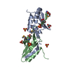 1mo1S S: Starting model for refinement |
|---|---|
| Similar structure data |
-Links
 PDBj
PDBj- Assembly
| Deposited unit | 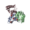
| |||||||||||||||||||||||||||||||||||||||||||||||||||||
|---|---|---|---|---|---|---|---|---|---|---|---|---|---|---|---|---|---|---|---|---|---|---|---|---|---|---|---|---|---|---|---|---|---|---|---|---|---|---|---|---|---|---|---|---|---|---|---|---|---|---|---|---|---|---|
| 1 | 
| |||||||||||||||||||||||||||||||||||||||||||||||||||||
| 2 | 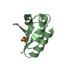
| |||||||||||||||||||||||||||||||||||||||||||||||||||||
| 3 | 
| |||||||||||||||||||||||||||||||||||||||||||||||||||||
| Unit cell |
| |||||||||||||||||||||||||||||||||||||||||||||||||||||
| Components on special symmetry positions |
| |||||||||||||||||||||||||||||||||||||||||||||||||||||
| Noncrystallographic symmetry (NCS) | NCS domain:
NCS domain segments: Component-ID: _ / Beg auth comp-ID: MET / Beg label comp-ID: MET / End auth comp-ID: GLY / End label comp-ID: GLY / Refine code: _ / Auth seq-ID: 1 - 84 / Label seq-ID: 1 - 84
NCS ensembles :
|
-Components
| #1: Protein | Mass: 9973.345 Da / Num. of mol.: 3 Source method: isolated from a genetically manipulated source Source: (gene. exp.) Ruminiclostridium cellulolyticum H10 (bacteria)Strain: H10 / Gene: Ccel_0806 / Production host: #2: Chemical | ChemComp-SO4 /   Mass: 96.063 Da / Num. of mol.: 7 / Source method: obtained synthetically / Formula: SO4 / Feature type: SUBJECT OF INVESTIGATION Mass: 96.063 Da / Num. of mol.: 7 / Source method: obtained synthetically / Formula: SO4 / Feature type: SUBJECT OF INVESTIGATION#3: Water | ChemComp-HOH / |  Mass: 18.015 Da / Num. of mol.: 69 / Source method: isolated from a natural source / Formula: H2O Mass: 18.015 Da / Num. of mol.: 69 / Source method: isolated from a natural source / Formula: H2OHas ligand of interest | Y | |
|---|
-Experimental details
-Experiment
| Experiment | Method: X-RAY DIFFRACTION / Number of used crystals: 1 |
|---|
- Sample preparation
| Crystal | Density Matthews: 3.46 Å3/Da / Density % sol: 64.44 % |
|---|---|
| Crystal grow | Temperature: 288 K / Method: vapor diffusion, hanging drop Details: 0.2 M Sodium sulfate, 20% w/v PEG3350, 10% w/v Ethylene glycol |
-Data collection
| Diffraction | Mean temperature: 100 K / Serial crystal experiment: N |
|---|---|
| Diffraction source | Source: SYNCHROTRON / Site: SSRF / Beamline: BL18U1 / Wavelength: 0.97915 Å |
| Detector | Type: DECTRIS PILATUS3 6M / Detector: PIXEL / Date: Jan 7, 2021 |
| Radiation | Protocol: SINGLE WAVELENGTH / Monochromatic (M) / Laue (L): M / Scattering type: x-ray |
| Radiation wavelength | Wavelength: 0.97915 Å / Relative weight: 1 |
| Reflection | Resolution: 2.37→30 Å / Num. obs: 17552 / % possible obs: 99.6 % / Redundancy: 1.9 % / CC1/2: 0.999 / Rmerge(I) obs: 0.015 / Net I/σ(I): 36.4 |
| Reflection shell | Resolution: 2.37→2.45 Å / Rmerge(I) obs: 0.061 / Num. unique obs: 1694 / CC1/2: 0.988 |
- Processing
| Software |
| |||||||||||||||||||||||||||||||||||
|---|---|---|---|---|---|---|---|---|---|---|---|---|---|---|---|---|---|---|---|---|---|---|---|---|---|---|---|---|---|---|---|---|---|---|---|---|
| Refinement | Method to determine structure: MOLECULAR REPLACEMENT Starting model: 1MO1 Resolution: 2.37→30 Å / Cor.coef. Fo:Fc: 0.942 / Cor.coef. Fo:Fc free: 0.895 / SU B: 6.137 / SU ML: 0.149 / Cross valid method: THROUGHOUT / σ(F): 0 / ESU R: 0.245 / ESU R Free: 0.213 / Stereochemistry target values: MAXIMUM LIKELIHOOD Details: HYDROGENS HAVE BEEN ADDED IN THE RIDING POSITIONS U VALUES : REFINED INDIVIDUALLY
| |||||||||||||||||||||||||||||||||||
| Solvent computation | Ion probe radii: 0.8 Å / Shrinkage radii: 0.8 Å / VDW probe radii: 1.2 Å / Solvent model: MASK | |||||||||||||||||||||||||||||||||||
| Displacement parameters | Biso max: 161.74 Å2 / Biso mean: 54.822 Å2 / Biso min: 23.89 Å2
| |||||||||||||||||||||||||||||||||||
| Refinement step | Cycle: final / Resolution: 2.37→30 Å
| |||||||||||||||||||||||||||||||||||
| Refine LS restraints NCS | Refine-ID: X-RAY DIFFRACTION / Type: interatomic distance / Weight position: 0.05
| |||||||||||||||||||||||||||||||||||
| LS refinement shell | Resolution: 2.37→2.43 Å / Rfactor Rfree error: 0
|
