 Movie
Movie Controller

[English] 日本語
 Yorodumi
- PDB-7fex: Crystal structure of Sus scrofa Schlafen11 N-terminal domain (2.69 A) -
+ Open data

- Basic information
| Entry | Database: PDB / ID: 7fex | ||||||
|---|---|---|---|---|---|---|---|
| Title | Crystal structure of Sus scrofa Schlafen11 N-terminal domain (2.69 A) | ||||||
 Components Components | SLFN11 | ||||||
 Keywords Keywords | ANTIVIRAL PROTEIN / Host antiviral protein / RNA helicase | ||||||
| Function / homology |  Function and homology information Function and homology informationreplication fork arrest / negative regulation of G1/S transition of mitotic cell cycle / site of DNA damage / defense response to virus / tRNA binding / chromatin remodeling / DNA damage response / ATP hydrolysis activity / nucleoplasm / ATP binding / cytosol Similarity search - Function | ||||||
| Biological species |  | ||||||
| Method |  X-RAY DIFFRACTION / SYNCHROTRON / SAD / Resolution: 2.69 Å X-RAY DIFFRACTION / SYNCHROTRON / SAD / Resolution: 2.69 Å | ||||||
 Authors Authors | Hou, P. / Hao, W. / Cui, S. | ||||||
 Citation Citation | Journal: Nucleic Acids Res. / Year: 2023 Title: Structural and biochemical characterization of Schlafen11 N-terminal domain. Authors: Hou, P. / Hao, W. / Qin, B. / Li, M. / Zhao, R. / Cui, S. | ||||||
| History |
|
- Structure visualization
| Structure viewer | Molecule: MolmilJmol/JSmol |
|---|
- Downloads & links
-Download
| PDBx/mmCIF format | 7fex.cif.gz | 144.8 KB | Display | PDBx/mmCIF format |
|---|---|---|---|---|
| PDB format | pdb7fex.ent.gz | 113.2 KB | Display | PDB format |
| PDBx/mmJSON format | 7fex.json.gz | Tree view | PDBx/mmJSON format | |
| Others |  Other downloads Other downloads |
-Validation report
| Arichive directory | https://data.pdbj.org/pub/pdb/validation_reports/fe/7fexftp://data.pdbj.org/pub/pdb/validation_reports/fe/7fex | HTTPS FTP |
|---|
-Related structure data
| Similar structure data |
|---|
-Links
 PDBj
PDBj- Assembly
| Deposited unit | 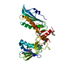
| ||||||||
|---|---|---|---|---|---|---|---|---|---|
| 1 |
| ||||||||
| Unit cell |
|
-Components
| #1: Protein | Mass: 38858.996 Da / Num. of mol.: 1 Source method: isolated from a genetically manipulated source Source: (gene. exp.)  |
|---|---|
| #2: Water | ChemComp-HOH /  Mass: 18.015 Da / Num. of mol.: 10 / Source method: isolated from a natural source / Formula: H2O Mass: 18.015 Da / Num. of mol.: 10 / Source method: isolated from a natural source / Formula: H2O |
| Has ligand of interest | N |
| Has protein modification | Y |
-Experimental details
-Experiment
| Experiment | Method: X-RAY DIFFRACTION / Number of used crystals: 1 |
|---|
- Sample preparation
| Crystal | Density Matthews: 2.7 Å3/Da / Density % sol: 54.42 % |
|---|---|
| Crystal grow | Temperature: 291 K / Method: vapor diffusion, hanging drop / pH: 7.5 Details: 0.1M HEPES pH7.5, 22%PEG3350, 0.15M Lithium sulfate |
-Data collection
| Diffraction | Mean temperature: 100 K / Serial crystal experiment: N |
|---|---|
| Diffraction source | Source: SYNCHROTRON / Site: SSRF  / Beamline: BL19U1 / Wavelength: 0.979 Å / Beamline: BL19U1 / Wavelength: 0.979 Å |
| Detector | Type: DECTRIS PILATUS 6M / Detector: PIXEL / Date: Oct 20, 2019 |
| Radiation | Protocol: SINGLE WAVELENGTH / Monochromatic (M) / Laue (L): M / Scattering type: x-ray |
| Radiation wavelength | Wavelength: 0.979 Å / Relative weight: 1 |
| Reflection | Resolution: 2.69→43.564 Å / Num. obs: 22498 / % possible obs: 99.9 % / Observed criterion σ(I): -3 / Redundancy: 34.4 % / Biso Wilson estimate: 61.86 Å2 / CC1/2: 0.999 / Rmerge(I) obs: 0.143 / Net I/σ(I): 26.33 |
| Reflection shell | Resolution: 2.69→2.71 Å / Redundancy: 34.8 % / Rmerge(I) obs: 1.15 / Num. unique obs: 3610 / CC1/2: 0.886 / % possible all: 99.8 |
- Processing
| Software |
| ||||||||||||||||||||||||||||||||||||||||||||||||||||||||||||
|---|---|---|---|---|---|---|---|---|---|---|---|---|---|---|---|---|---|---|---|---|---|---|---|---|---|---|---|---|---|---|---|---|---|---|---|---|---|---|---|---|---|---|---|---|---|---|---|---|---|---|---|---|---|---|---|---|---|---|---|---|---|
| Refinement | Method to determine structure: SAD / Resolution: 2.69→43.56 Å / SU ML: 0.4 / Cross valid method: THROUGHOUT / σ(F): 1.34 / Phase error: 26.41 / Stereochemistry target values: ML
| ||||||||||||||||||||||||||||||||||||||||||||||||||||||||||||
| Solvent computation | Shrinkage radii: 0.9 Å / VDW probe radii: 1.11 Å / Solvent model: FLAT BULK SOLVENT MODEL | ||||||||||||||||||||||||||||||||||||||||||||||||||||||||||||
| Displacement parameters | Biso max: 166.51 Å2 / Biso mean: 66.8739 Å2 / Biso min: 36.12 Å2 | ||||||||||||||||||||||||||||||||||||||||||||||||||||||||||||
| Refinement step | Cycle: final / Resolution: 2.69→43.56 Å
| ||||||||||||||||||||||||||||||||||||||||||||||||||||||||||||
| LS refinement shell | Refine-ID: X-RAY DIFFRACTION / Rfactor Rfree error: 0 / Total num. of bins used: 9 / % reflection obs: 100 %
| ||||||||||||||||||||||||||||||||||||||||||||||||||||||||||||
| Refinement TLS params. | Method: refined / Origin x: 2.3292 Å / Origin y: 15.9678 Å / Origin z: -9.8295 Å
| ||||||||||||||||||||||||||||||||||||||||||||||||||||||||||||
| Refinement TLS group | Selection details: Chain A |
