 Movie
Movie Controller

[English] 日本語
 Yorodumi
- PDB-7fbo: geranyl pyrophosphate C6-methyltransferase BezA binding with S-ad... -
+ Open data

- Basic information
| Entry | Database: PDB / ID: 7fbo | ||||||
|---|---|---|---|---|---|---|---|
| Title | geranyl pyrophosphate C6-methyltransferase BezA binding with S-adenosylhomocysteine | ||||||
 Components Components | BezA | ||||||
 Keywords Keywords | TRANSFERASE / methyltransferase geranyl pyrophosphate S-adenosylhomocysteine | ||||||
| Function / homology | S-ADENOSYL-L-HOMOCYSTEINE Function and homology information Function and homology information | ||||||
| Biological species |  Streptomyces sp. RI-18-2 (bacteria) Streptomyces sp. RI-18-2 (bacteria) | ||||||
| Method |  X-RAY DIFFRACTION / SYNCHROTRON / MOLECULAR REPLACEMENT / Resolution: 2.56 Å X-RAY DIFFRACTION / SYNCHROTRON / MOLECULAR REPLACEMENT / Resolution: 2.56 Å | ||||||
 Authors Authors | Tsutsumi, H. / Moriwaki, Y. / Terada, T. / Shimizu, K. / Katsuyama, Y. / Ohnishi, Y. | ||||||
| Funding support |  Japan, 1items Japan, 1items
| ||||||
 Citation Citation | Journal: Angew.Chem.Int.Ed.Engl. / Year: 2022 Title: Structural and Molecular Basis of the Catalytic Mechanism of Geranyl Pyrophosphate C6-Methyltransferase: Creation of an Unprecedented Farnesyl Pyrophosphate C6-Methyltransferase. Authors: Tsutsumi, H. / Moriwaki, Y. / Terada, T. / Shimizu, K. / Shin-Ya, K. / Katsuyama, Y. / Ohnishi, Y. #1: Journal: J Am Chem Soc / Year: 2018 Title: Unprecedented Cyclization Catalyzed by a Cytochrome P450 in Benzastatin Biosynthesis. Authors: Tsutsumi, H. / Katsuyama, Y. / Izumikawa, M. / Takagi, M. / Fujie, M. / Satoh, N. / Shin-Ya, K. / Ohnishi, Y. | ||||||
| History |
|
- Structure visualization
| Structure viewer | Molecule: MolmilJmol/JSmol |
|---|
- Downloads & links
-Download
| PDBx/mmCIF format | 7fbo.cif.gz | 222.6 KB | Display | PDBx/mmCIF format |
|---|---|---|---|---|
| PDB format | pdb7fbo.ent.gz | 143.7 KB | Display | PDB format |
| PDBx/mmJSON format | 7fbo.json.gz | Tree view | PDBx/mmJSON format | |
| Others |  Other downloads Other downloads |
-Validation report
| Arichive directory | https://data.pdbj.org/pub/pdb/validation_reports/fb/7fboftp://data.pdbj.org/pub/pdb/validation_reports/fb/7fbo | HTTPS FTP |
|---|
-Related structure data
| Related structure data |  7fbhSC S: Starting model for refinement C: citing same article ( |
|---|---|
| Similar structure data |
-Links
 PDBj
PDBj- Assembly
| Deposited unit | 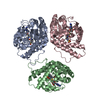
| ||||||||||||
|---|---|---|---|---|---|---|---|---|---|---|---|---|---|
| 1 | 
| ||||||||||||
| Unit cell |
| ||||||||||||
| Components on special symmetry positions |
|
-Components
| #1: Protein | Mass: 33605.676 Da / Num. of mol.: 3 Source method: isolated from a genetically manipulated source Source: (gene. exp.) Streptomyces sp. RI-18-2 (bacteria) / Plasmid: pColdI / Production host: #2: Chemical |   Mass: 238.305 Da / Num. of mol.: 3 / Source method: obtained synthetically / Formula: C8H18N2O4S / Feature type: SUBJECT OF INVESTIGATION / Comment: pH buffer*YM Mass: 238.305 Da / Num. of mol.: 3 / Source method: obtained synthetically / Formula: C8H18N2O4S / Feature type: SUBJECT OF INVESTIGATION / Comment: pH buffer*YM#3: Chemical |   Type: L-peptide linking / Mass: 384.411 Da / Num. of mol.: 3 / Source method: obtained synthetically / Formula: C14H20N6O5S / Feature type: SUBJECT OF INVESTIGATION Type: L-peptide linking / Mass: 384.411 Da / Num. of mol.: 3 / Source method: obtained synthetically / Formula: C14H20N6O5S / Feature type: SUBJECT OF INVESTIGATION#4: Water | ChemComp-HOH / |  Mass: 18.015 Da / Num. of mol.: 197 / Source method: isolated from a natural source / Formula: H2O Mass: 18.015 Da / Num. of mol.: 197 / Source method: isolated from a natural source / Formula: H2OHas ligand of interest | Y | |
|---|
-Experimental details
-Experiment
| Experiment | Method: X-RAY DIFFRACTION / Number of used crystals: 1 |
|---|
- Sample preparation
| Crystal | Density Matthews: 2.34 Å3/Da / Density % sol: 47.48 % |
|---|---|
| Crystal grow | Temperature: 293 K / Method: vapor diffusion, hanging drop Details: 0.1 M HEPES (pH 7.5), 25% PEG3350, 0.2 M CH3COONH4. 0.01 M spermine tetrahydrochloride Soaking using 0.1 M HEPES (pH7.5), 35% PEG3350, 1.0 mM SAH. |
-Data collection
| Diffraction | Mean temperature: 95 K / Serial crystal experiment: N |
|---|---|
| Diffraction source | Source: SYNCHROTRON / Site: Photon Factory / Beamline: BL-1A / Wavelength: 1 Å |
| Detector | Type: DECTRIS EIGER X 4M / Detector: PIXEL / Date: Jun 3, 2019 |
| Radiation | Protocol: SINGLE WAVELENGTH / Monochromatic (M) / Laue (L): M / Scattering type: x-ray |
| Radiation wavelength | Wavelength: 1 Å / Relative weight: 1 |
| Reflection | Resolution: 2.56→50.34 Å / Num. obs: 30941 / % possible obs: 100 % / Redundancy: 7.5 % / Biso Wilson estimate: 40.1 Å2 / CC1/2: 0.996 / Rmerge(I) obs: 0.15 / Rpim(I) all: 0.055 / Rrim(I) all: 0.15 / Net I/σ(I): 11.2 |
| Reflection shell | Resolution: 2.56→2.67 Å / Redundancy: 7.8 % / Rmerge(I) obs: 0.864 / Mean I/σ(I) obs: 2.5 / Num. unique obs: 3824 / CC1/2: 0.835 / Rpim(I) all: 0.33 / Rrim(I) all: 0.926 / % possible all: 100 |
- Processing
| Software |
| |||||||||||||||||||||||||||||||||||||||||||||||||||||||||||||||||||||||||||||||||||||||||||||||||||||||||
|---|---|---|---|---|---|---|---|---|---|---|---|---|---|---|---|---|---|---|---|---|---|---|---|---|---|---|---|---|---|---|---|---|---|---|---|---|---|---|---|---|---|---|---|---|---|---|---|---|---|---|---|---|---|---|---|---|---|---|---|---|---|---|---|---|---|---|---|---|---|---|---|---|---|---|---|---|---|---|---|---|---|---|---|---|---|---|---|---|---|---|---|---|---|---|---|---|---|---|---|---|---|---|---|---|---|---|
| Refinement | Method to determine structure: MOLECULAR REPLACEMENT Starting model: 7FBH Resolution: 2.56→50.34 Å / SU ML: 0.3289 / Cross valid method: FREE R-VALUE / σ(F): 0 / Phase error: 26.0371 Stereochemistry target values: GeoStd + Monomer Library + CDL v1.2
| |||||||||||||||||||||||||||||||||||||||||||||||||||||||||||||||||||||||||||||||||||||||||||||||||||||||||
| Solvent computation | Shrinkage radii: 0.9 Å / VDW probe radii: 1.11 Å / Solvent model: FLAT BULK SOLVENT MODEL | |||||||||||||||||||||||||||||||||||||||||||||||||||||||||||||||||||||||||||||||||||||||||||||||||||||||||
| Displacement parameters | Biso mean: 44.08 Å2 | |||||||||||||||||||||||||||||||||||||||||||||||||||||||||||||||||||||||||||||||||||||||||||||||||||||||||
| Refinement step | Cycle: LAST / Resolution: 2.56→50.34 Å
| |||||||||||||||||||||||||||||||||||||||||||||||||||||||||||||||||||||||||||||||||||||||||||||||||||||||||
| Refine LS restraints |
| |||||||||||||||||||||||||||||||||||||||||||||||||||||||||||||||||||||||||||||||||||||||||||||||||||||||||
| LS refinement shell |
|
