 Movie
Movie Controller

[English] 日本語
 Yorodumi
- PDB-7fa0: The crystal structure of VyPAL2-C214A, a dead mutant of VyPAL2 pe... -
+ Open data

- Basic information
| Entry | Database: PDB / ID: 7fa0 | |||||||||
|---|---|---|---|---|---|---|---|---|---|---|
| Title | The crystal structure of VyPAL2-C214A, a dead mutant of VyPAL2 peptide asparaginyl ligase in form II | |||||||||
 Components Components | Peptide Asparaginyl Ligases | |||||||||
 Keywords Keywords | PLANT PROTEIN / AEP / PAL / Legumain / peptide ligase | |||||||||
| Function / homology |  Function and homology information Function and homology informationvacuolar protein processing / vacuole / : / cysteine-type endopeptidase activity Similarity search - Function | |||||||||
| Biological species |  Viola philippica (plant) Viola philippica (plant) | |||||||||
| Method |  X-RAY DIFFRACTION / SYNCHROTRON / MOLECULAR REPLACEMENT / Resolution: 1.8 Å X-RAY DIFFRACTION / SYNCHROTRON / MOLECULAR REPLACEMENT / Resolution: 1.8 Å | |||||||||
 Authors Authors | Hu, S. / Sahili, A. / Lescar, J. | |||||||||
| Funding support |  Singapore, 1items Singapore, 1items
| |||||||||
 Citation Citation | Journal: Plant Cell / Year: 2022 Title: Structural basis for proenzyme maturation, substrate recognition, and ligation by a hyperactive peptide asparaginyl ligase. Authors: Hu, S. / El Sahili, A. / Kishore, S. / Wong, Y.H. / Hemu, X. / Goh, B.C. / Zhipei, S. / Wang, Z. / Tam, J.P. / Liu, C.F. / Lescar, J. | |||||||||
| History |
|
- Structure visualization
| Structure viewer | Molecule: MolmilJmol/JSmol |
|---|
- Downloads & links
-Download
| PDBx/mmCIF format | 7fa0.cif.gz | 241.4 KB | Display | PDBx/mmCIF format |
|---|---|---|---|---|
| PDB format | pdb7fa0.ent.gz | 192.8 KB | Display | PDB format |
| PDBx/mmJSON format | 7fa0.json.gz | Tree view | PDBx/mmJSON format | |
| Others |  Other downloads Other downloads |
-Validation report
| Arichive directory | https://data.pdbj.org/pub/pdb/validation_reports/fa/7fa0ftp://data.pdbj.org/pub/pdb/validation_reports/fa/7fa0 | HTTPS FTP |
|---|
-Related structure data
| Related structure data |  7f5jC  7f5pC 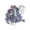 7f5qC  6idvS C: citing same article ( S: Starting model for refinement |
|---|---|
| Similar structure data |
-Links
 PDBj
PDBj
- Assembly
| Deposited unit | 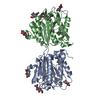
| ||||||||
|---|---|---|---|---|---|---|---|---|---|
| 1 | 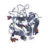
| ||||||||
| 2 | 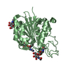
| ||||||||
| Unit cell |
|
-Components
| #1: Protein | Mass: 31100.783 Da / Num. of mol.: 2 / Mutation: C214A Source method: isolated from a genetically manipulated source Details: D171 (forming SNN), residue 172 is HD0 (HIS + SNN) / Source: (gene. exp.) Viola philippica (plant) / Production host:   Spodoptera frugiperda (fall armyworm) / References: UniProt: A0A509GV09 Spodoptera frugiperda (fall armyworm) / References: UniProt: A0A509GV09#2: Polysaccharide | alpha-L-fucopyranose-(1-6)-2-acetamido-2-deoxy-beta-D-glucopyranose | Source method: isolated from a genetically manipulated source #3: Polysaccharide | 2-acetamido-2-deoxy-beta-D-glucopyranose-(1-4)-2-acetamido-2-deoxy-beta-D-glucopyranose | Source method: isolated from a genetically manipulated source #4: Sugar | ChemComp-NAG /   Type: D-saccharide, beta linking / Mass: 221.208 Da / Num. of mol.: 4 / Source method: obtained synthetically / Formula: C8H15NO6 Type: D-saccharide, beta linking / Mass: 221.208 Da / Num. of mol.: 4 / Source method: obtained synthetically / Formula: C8H15NO6#5: Water | ChemComp-HOH / |  Mass: 18.015 Da / Num. of mol.: 471 / Source method: isolated from a natural source / Formula: H2O Mass: 18.015 Da / Num. of mol.: 471 / Source method: isolated from a natural source / Formula: H2OHas ligand of interest | N | |
|---|
-Experimental details
-Experiment
| Experiment | Method: X-RAY DIFFRACTION / Number of used crystals: 1 |
|---|
- Sample preparation
| Crystal | Density Matthews: 2.24 Å3/Da / Density % sol: 45.04 % |
|---|---|
| Crystal grow | Temperature: 293.15 K / Method: vapor diffusion, sitting drop / Details: 0.2 M Potassium formate, 20% PEG 3350 |
-Data collection
| Diffraction | Mean temperature: 100 K / Serial crystal experiment: N |
|---|---|
| Diffraction source | Source: SYNCHROTRON / Site: SLS  / Beamline: X06SA / Wavelength: 1.00001 Å / Beamline: X06SA / Wavelength: 1.00001 Å |
| Detector | Type: DECTRIS EIGER X 16M / Detector: PIXEL / Date: Jun 3, 2021 |
| Radiation | Protocol: SINGLE WAVELENGTH / Monochromatic (M) / Laue (L): M / Scattering type: x-ray |
| Radiation wavelength | Wavelength: 1.00001 Å / Relative weight: 1 |
| Reflection | Resolution: 1.8→46.33 Å / Num. obs: 51971 / % possible obs: 99.09 % / Redundancy: 6.7 % / Biso Wilson estimate: 27.28 Å2 / CC1/2: 0.997 / Rmerge(I) obs: 0.09147 / Rpim(I) all: 0.03844 / Rrim(I) all: 0.09946 / Net I/σ(I): 12.65 |
| Reflection shell | Resolution: 1.8→1.864 Å / Redundancy: 6.2 % / Rmerge(I) obs: 1.008 / Mean I/σ(I) obs: 2.12 / Num. unique obs: 5111 / CC1/2: 0.698 / CC star: 0.907 / Rpim(I) all: 0.4336 / Rrim(I) all: 1.1 / % possible all: 98.35 |
- Processing
| Software |
| |||||||||||||||||||||||||||||||||||||||||||||||||||||||||||||||||||||||||||
|---|---|---|---|---|---|---|---|---|---|---|---|---|---|---|---|---|---|---|---|---|---|---|---|---|---|---|---|---|---|---|---|---|---|---|---|---|---|---|---|---|---|---|---|---|---|---|---|---|---|---|---|---|---|---|---|---|---|---|---|---|---|---|---|---|---|---|---|---|---|---|---|---|---|---|---|---|
| Refinement | Method to determine structure: MOLECULAR REPLACEMENT Starting model: 6IDV Resolution: 1.8→46.33 Å / Cor.coef. Fo:Fc: 0.965 / Cor.coef. Fo:Fc free: 0.953 / SU R Cruickshank DPI: 0.117 / Cross valid method: THROUGHOUT / SU R Blow DPI: 0.125 / SU Rfree Blow DPI: 0.111 / SU Rfree Cruickshank DPI: 0.107
| |||||||||||||||||||||||||||||||||||||||||||||||||||||||||||||||||||||||||||
| Displacement parameters | Biso mean: 33.24 Å2
| |||||||||||||||||||||||||||||||||||||||||||||||||||||||||||||||||||||||||||
| Refine analyze | Luzzati coordinate error obs: 0.21 Å | |||||||||||||||||||||||||||||||||||||||||||||||||||||||||||||||||||||||||||
| Refinement step | Cycle: LAST / Resolution: 1.8→46.33 Å
| |||||||||||||||||||||||||||||||||||||||||||||||||||||||||||||||||||||||||||
| Refine LS restraints |
| |||||||||||||||||||||||||||||||||||||||||||||||||||||||||||||||||||||||||||
| LS refinement shell | Resolution: 1.8→1.81 Å
| |||||||||||||||||||||||||||||||||||||||||||||||||||||||||||||||||||||||||||
| Refinement TLS params. | Refine-ID: X-RAY DIFFRACTION
| |||||||||||||||||||||||||||||||||||||||||||||||||||||||||||||||||||||||||||
| Refinement TLS group |
|
