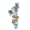
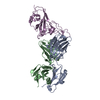
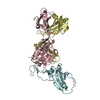
Entry Database : PDB / ID : 7f7hTitle SARS-CoV-2 S protein RBD in complex with A8-1 Fab Heavy chain of A8-1 Fab Light chain of A8-1 Fab Spike glycoprotein S1 Keywords / / / / Function / homology Function Domain/homology Component
/ / / / / / / / / / / / / / / / / / / / / / / / / / / / / / / / / / / / / / / / / / / / / / / / / / / / / / / / / / / / / / / / / Biological species Homo sapiens (human)Method / / / Resolution : 3.19 Å Authors Dou, Y. / Wang, X. / Wang, K. / Liu, P. / Lu, B. Journal : To Be Published Title : High throughput isolation of potent neutralizing antibodies from convalescent COVID-19 patients.Authors : Dou, Y. / Jia, Z. / Deng, Y. / Lan, J. / Liu, H. History Deposition Jun 29, 2021 Deposition site / Processing site Revision 1.0 Jun 15, 2022 Provider / Type Revision 1.1 Nov 29, 2023 Group / Refinement descriptionCategory chem_comp_atom / chem_comp_bond ... chem_comp_atom / chem_comp_bond / pdbx_initial_refinement_model / struct_ncs_dom_lim Item _struct_ncs_dom_lim.beg_auth_comp_id / _struct_ncs_dom_lim.beg_label_asym_id ... _struct_ncs_dom_lim.beg_auth_comp_id / _struct_ncs_dom_lim.beg_label_asym_id / _struct_ncs_dom_lim.beg_label_comp_id / _struct_ncs_dom_lim.beg_label_seq_id / _struct_ncs_dom_lim.end_auth_comp_id / _struct_ncs_dom_lim.end_label_asym_id / _struct_ncs_dom_lim.end_label_comp_id / _struct_ncs_dom_lim.end_label_seq_id
Show all Show less
 Movie
Movie Controller


 Open data
 Basic information
 Components
 Keywords
 Function and homology information
 Homo sapiens (human)
 Severe acute respiratory syndrome coronavirus 2
 X-RAY DIFFRACTION /
  Authors
 Citation
 Structure visualization
 Downloads & links
 Other downloads
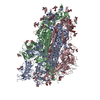
 PDBj
PDBj


 Assembly