 Movie
Movie Controller

[English] 日本語
 Yorodumi
- PDB-7f7f: Cryo-EM structure of Dnf1 from Saccharomyces cerevisiae in yeast ... -
+ Open data

- Basic information
| Entry | Database: PDB / ID: 7f7f | ||||||||||||||||||||||||||||||||||||
|---|---|---|---|---|---|---|---|---|---|---|---|---|---|---|---|---|---|---|---|---|---|---|---|---|---|---|---|---|---|---|---|---|---|---|---|---|---|
| Title | Cryo-EM structure of Dnf1 from Saccharomyces cerevisiae in yeast lipids with beryllium fluoride (resting state) | ||||||||||||||||||||||||||||||||||||
 Components Components |
| ||||||||||||||||||||||||||||||||||||
 Keywords Keywords | LIPID TRANSPORT / P4-ATPases | ||||||||||||||||||||||||||||||||||||
| Function / homology |  Function and homology information Function and homology informationregulation of vacuole organization / glycosylceramide flippase activity / mating projection tip membrane / aminophospholipid translocation / phosphatidylcholine flippase activity / Ion transport by P-type ATPases / phosphatidylserine flippase activity / phospholipid-translocating ATPase complex / ceramide translocation / phosphatidylserine floppase activity ...regulation of vacuole organization / glycosylceramide flippase activity / mating projection tip membrane / aminophospholipid translocation / phosphatidylcholine flippase activity / Ion transport by P-type ATPases / phosphatidylserine flippase activity / phospholipid-translocating ATPase complex / ceramide translocation / phosphatidylserine floppase activity / ATPase-coupled intramembrane lipid carrier activity / phosphatidylethanolamine flippase activity / phosphatidylcholine floppase activity / cell septum / cellular bud neck / P-type phospholipid transporter / phospholipid translocation / establishment or maintenance of cell polarity / Neutrophil degranulation / cell periphery / intracellular protein transport / endocytosis / cell surface receptor signaling pathway / endosome membrane / Golgi apparatus / magnesium ion binding / endoplasmic reticulum / ATP hydrolysis activity / mitochondrion / ATP binding / identical protein binding / plasma membrane / cytoplasm Similarity search - Function | ||||||||||||||||||||||||||||||||||||
| Biological species |  | ||||||||||||||||||||||||||||||||||||
| Method | ELECTRON MICROSCOPY / single particle reconstruction / cryo EM / Resolution: 3.81 Å | ||||||||||||||||||||||||||||||||||||
 Authors Authors | Xu, J. / He, Y. / Wu, X. / Li, L. | ||||||||||||||||||||||||||||||||||||
| Funding support |  China, 1items China, 1items
| ||||||||||||||||||||||||||||||||||||
 Citation Citation | Journal: Cell Rep / Year: 2022 Title: Conformational changes of a phosphatidylcholine flippase in lipid membranes. Authors: Jinkun Xu / Yilin He / Xiaofei Wu / Long Li / Abstract: Type 4 P-type ATPases (P4-ATPases) actively and selectively translocate phospholipids across membrane bilayers. Driven by ATP hydrolysis, P4-ATPases undergo conformational changes during lipid ...Type 4 P-type ATPases (P4-ATPases) actively and selectively translocate phospholipids across membrane bilayers. Driven by ATP hydrolysis, P4-ATPases undergo conformational changes during lipid flipping. It is unclear how the active flipping states of P4-ATPases are regulated in the lipid membranes, especially for phosphatidylcholine (PC)-flipping P4-ATPases whose substrate, PC, is a substantial component of membranes. Here, we report the cryoelectron microscopy structures of a yeast PC-flipping P4-ATPase, Dnf1, in lipid environments. In native yeast lipids, Dnf1 adopts a conformation in which the lipid flipping pathway is disrupted. Only when the lipid composition is changed can Dnf1 be captured in the active conformations that enable lipid flipping. These results suggest that, in the native membrane, Dnf1 may stay in an idle conformation that is unable to support the trans-membrane movement of lipids. Dnf1 may have altered conformational preferences in membranes with different lipid compositions. | ||||||||||||||||||||||||||||||||||||
| History |
|
- Structure visualization
| Structure viewer | Molecule: MolmilJmol/JSmol |
|---|
- Downloads & links
-Download
| PDBx/mmCIF format | 7f7f.cif.gz | 287.3 KB | Display | PDBx/mmCIF format |
|---|---|---|---|---|
| PDB format | pdb7f7f.ent.gz | 221.1 KB | Display | PDB format |
| PDBx/mmJSON format | 7f7f.json.gz | Tree view | PDBx/mmJSON format | |
| Others |  Other downloads Other downloads |
-Validation report
| Arichive directory | https://data.pdbj.org/pub/pdb/validation_reports/f7/7f7fftp://data.pdbj.org/pub/pdb/validation_reports/f7/7f7f | HTTPS FTP |
|---|
-Related structure data
| Related structure data |  31487MC 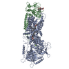 7drxC  7dshC 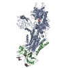 7dsiC  7whvC 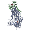 7whwC M: map data used to model this data C: citing same article ( |
|---|---|
| Similar structure data |


-Links
 PDBj
PDBj


- Assembly
| Deposited unit | 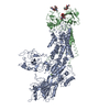
|
|---|---|
| 1 |
|
-Components
-Protein , 2 types, 2 molecules AB
| #1: Protein | Mass: 178000.172 Da / Num. of mol.: 1 Source method: isolated from a genetically manipulated source Source: (gene. exp.) References: UniProt: P32660, P-type phospholipid transporter |
|---|---|
| #2: Protein | Mass: 47490.395 Da / Num. of mol.: 1 Source method: isolated from a genetically manipulated source Source: (gene. exp.) |
-Sugars , 3 types, 3 molecules
| #3: Polysaccharide | 2-acetamido-2-deoxy-beta-D-glucopyranose-(1-4)-2-acetamido-2-deoxy-beta-D-glucopyranose Source method: isolated from a genetically manipulated source |
|---|---|
| #4: Polysaccharide | beta-D-mannopyranose-(1-4)-2-acetamido-2-deoxy-beta-D-glucopyranose-(1-4)-2-acetamido-2-deoxy-beta- ...beta-D-mannopyranose-(1-4)-2-acetamido-2-deoxy-beta-D-glucopyranose-(1-4)-2-acetamido-2-deoxy-beta-D-glucopyranose Source method: isolated from a genetically manipulated source |
| #5: Polysaccharide | 2-acetamido-2-deoxy-beta-D-glucopyranose-(1-6)-2-acetamido-2-deoxy-beta-D-glucopyranose Source method: isolated from a genetically manipulated source |
-Non-polymers , 1 types, 1 molecules 
| #6: Chemical | ChemComp-MG /  Mass: 24.305 Da / Num. of mol.: 1 Mass: 24.305 Da / Num. of mol.: 1Source method: isolated from a genetically manipulated source Formula: Mg |
|---|
-Details
| Has ligand of interest | N |
|---|---|
| Has protein modification | Y |
-Experimental details
-Experiment
| Experiment | Method: ELECTRON MICROSCOPY |
|---|---|
| EM experiment | Aggregation state: PARTICLE / 3D reconstruction method: single particle reconstruction |
- Sample preparation
| Component | Name: Dnf1_Lem3 complex / Type: COMPLEX / Entity ID: #1-#2 / Source: RECOMBINANT |
|---|---|
| Source (natural) | Organism: |
| Source (recombinant) | Organism: |
| Buffer solution | pH: 7.5 |
| Specimen | Embedding applied: NO / Shadowing applied: NO / Staining applied: NO / Vitrification applied: YES |
| Vitrification | Cryogen name: NITROGEN |
- Electron microscopy imaging
| Experimental equipment |  Model: Titan Krios / Image courtesy: FEI Company |
|---|---|
| Microscopy | Model: FEI TITAN KRIOS |
| Electron gun | Electron source:  FIELD EMISSION GUN / Accelerating voltage: 300 kV / Illumination mode: SPOT SCAN FIELD EMISSION GUN / Accelerating voltage: 300 kV / Illumination mode: SPOT SCAN |
| Electron lens | Mode: BRIGHT FIELD / Nominal defocus max: 2000 nm / Nominal defocus min: 1000 nm |
| Image recording | Electron dose: 8 e/Å2 / Film or detector model: DIRECT ELECTRON DE-16 (4k x 4k) |
- Processing
| Software | Name: PHENIX / Classification: refinement | ||||||||||||||||||||||||
|---|---|---|---|---|---|---|---|---|---|---|---|---|---|---|---|---|---|---|---|---|---|---|---|---|---|
| EM software | Name: PHENIX / Category: model refinement | ||||||||||||||||||||||||
| CTF correction | Type: PHASE FLIPPING AND AMPLITUDE CORRECTION | ||||||||||||||||||||||||
| 3D reconstruction | Resolution: 3.81 Å / Resolution method: FSC 0.143 CUT-OFF / Num. of particles: 212294 / Symmetry type: POINT | ||||||||||||||||||||||||
| Refine LS restraints |
|
