 Movie
Movie Controller

[English] 日本語
 Yorodumi
- PDB-7f50: X-ray crystal structure of Y149A mutated Hsp72-NBD in complex wit... -
+ Open data

- Basic information
| Entry | Database: PDB / ID: 7f50 | ||||||
|---|---|---|---|---|---|---|---|
| Title | X-ray crystal structure of Y149A mutated Hsp72-NBD in complex with AMPPnP | ||||||
 Components Components | Heat shock 70 kDa protein 1B | ||||||
 Keywords Keywords | HYDROLASE / Complex / Chaperone / nuclueotide-binding domain | ||||||
| Function / homology |  Function and homology information Function and homology informationcellular heat acclimation / negative regulation of inclusion body assembly / C3HC4-type RING finger domain binding / positive regulation of nucleotide-binding oligomerization domain containing 2 signaling pathway / positive regulation of microtubule nucleation / ATP-dependent protein disaggregase activity / positive regulation of tumor necrosis factor-mediated signaling pathway / regulation of mitotic spindle assembly / aggresome / cellular response to steroid hormone stimulus ...cellular heat acclimation / negative regulation of inclusion body assembly / C3HC4-type RING finger domain binding / positive regulation of nucleotide-binding oligomerization domain containing 2 signaling pathway / positive regulation of microtubule nucleation / ATP-dependent protein disaggregase activity / positive regulation of tumor necrosis factor-mediated signaling pathway / regulation of mitotic spindle assembly / aggresome / cellular response to steroid hormone stimulus / mRNA catabolic process / regulation of protein ubiquitination / HSF1-dependent transactivation / Regulation of HSF1-mediated heat shock response / negative regulation of extrinsic apoptotic signaling pathway in absence of ligand / Mitochondrial unfolded protein response (UPRmt) / Attenuation phase / ATP metabolic process / heat shock protein binding / inclusion body / protein folding chaperone / centriole / negative regulation of protein ubiquitination / HSP90 chaperone cycle for steroid hormone receptors (SHR) in the presence of ligand / positive regulation of erythrocyte differentiation / positive regulation of interleukin-8 production / ATP-dependent protein folding chaperone / : / negative regulation of cell growth / G protein-coupled receptor binding / PKR-mediated signaling / histone deacetylase binding / unfolded protein binding / positive regulation of proteasomal ubiquitin-dependent protein catabolic process / cellular response to heat / virus receptor activity / protein refolding / cellular response to oxidative stress / blood microparticle / vesicle / ficolin-1-rich granule lumen / protein stabilization / nuclear speck / ribonucleoprotein complex / signaling receptor binding / negative regulation of cell population proliferation / focal adhesion / ubiquitin protein ligase binding / Neutrophil degranulation / centrosome / positive regulation of gene expression / negative regulation of apoptotic process / perinuclear region of cytoplasm / enzyme binding / endoplasmic reticulum / ATP hydrolysis activity / protein-containing complex / mitochondrion / RNA binding / extracellular exosome / extracellular region / nucleoplasm / ATP binding / nucleus / plasma membrane / cytosol / cytoplasm Similarity search - Function | ||||||
| Biological species |  Homo sapiens (human) Homo sapiens (human) | ||||||
| Method |  X-RAY DIFFRACTION / SYNCHROTRON / MOLECULAR REPLACEMENT / Resolution: 1.703 Å X-RAY DIFFRACTION / SYNCHROTRON / MOLECULAR REPLACEMENT / Resolution: 1.703 Å | ||||||
 Authors Authors | Yokoyama, T. / Fujii, S. / Nabeshima, Y. / Mizuguchi, M. | ||||||
 Citation Citation | Journal: Iucrj / Year: 2022 Title: Neutron crystallographic analysis of the nucleotide-binding domain of Hsp72 in complex with ADP. Authors: Yokoyama, T. / Fujii, S. / Ostermann, A. / Schrader, T.E. / Nabeshima, Y. / Mizuguchi, M. | ||||||
| History |
|
- Structure visualization
| Structure viewer | Molecule: MolmilJmol/JSmol |
|---|
- Downloads & links
-Download
| PDBx/mmCIF format | 7f50.cif.gz | 98.8 KB | Display | PDBx/mmCIF format |
|---|---|---|---|---|
| PDB format | pdb7f50.ent.gz | 71 KB | Display | PDB format |
| PDBx/mmJSON format | 7f50.json.gz | Tree view | PDBx/mmJSON format | |
| Others |  Other downloads Other downloads |
-Validation report
| Arichive directory | https://data.pdbj.org/pub/pdb/validation_reports/f5/7f50ftp://data.pdbj.org/pub/pdb/validation_reports/f5/7f50 | HTTPS FTP |
|---|
-Related structure data
| Related structure data |  7f4xC 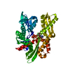 7f4zC 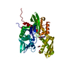 5aqzS S: Starting model for refinement C: citing same article ( |
|---|---|
| Similar structure data |
-Links
 PDBj
PDBj


- Assembly
| Deposited unit | 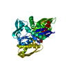
| ||||||||
|---|---|---|---|---|---|---|---|---|---|
| 1 |
| ||||||||
| Unit cell |
|
-Components
| #1: Protein | Mass: 42673.188 Da / Num. of mol.: 1 Source method: isolated from a genetically manipulated source Source: (gene. exp.) Homo sapiens (human) / Gene: HSPA1B, HSP72 / Production host:  | ||||||||
|---|---|---|---|---|---|---|---|---|---|
| #2: Chemical |   Mass: 24.305 Da / Num. of mol.: 2 / Source method: obtained synthetically / Formula: Mg / Feature type: SUBJECT OF INVESTIGATION Mass: 24.305 Da / Num. of mol.: 2 / Source method: obtained synthetically / Formula: Mg / Feature type: SUBJECT OF INVESTIGATION#3: Chemical | ChemComp-CL / |   Mass: 35.453 Da / Num. of mol.: 1 / Source method: obtained synthetically / Formula: Cl / Feature type: SUBJECT OF INVESTIGATION Mass: 35.453 Da / Num. of mol.: 1 / Source method: obtained synthetically / Formula: Cl / Feature type: SUBJECT OF INVESTIGATION#4: Chemical | ChemComp-ANP / |   Mass: 506.196 Da / Num. of mol.: 1 / Source method: obtained synthetically / Formula: C10H17N6O12P3 / Feature type: SUBJECT OF INVESTIGATION / Comment: AMP-PNP, energy-carrying molecule analogue*YM Mass: 506.196 Da / Num. of mol.: 1 / Source method: obtained synthetically / Formula: C10H17N6O12P3 / Feature type: SUBJECT OF INVESTIGATION / Comment: AMP-PNP, energy-carrying molecule analogue*YM#5: Water | ChemComp-HOH / |  Mass: 18.015 Da / Num. of mol.: 328 / Source method: isolated from a natural source / Formula: H2O Mass: 18.015 Da / Num. of mol.: 328 / Source method: isolated from a natural source / Formula: H2OHas ligand of interest | Y | |
-Experimental details
-Experiment
| Experiment | Method: X-RAY DIFFRACTION / Number of used crystals: 1 |
|---|
- Sample preparation
| Crystal | Density Matthews: 2.38 Å3/Da / Density % sol: 48.22 % |
|---|---|
| Crystal grow | Temperature: 293 K / Method: vapor diffusion, hanging drop Details: 29% PEG550 MME, 200 mM MgCl2 and 100 mM Tris-HCl pH 7.0 |
-Data collection
| Diffraction | Mean temperature: 100 K / Serial crystal experiment: N |
|---|---|
| Diffraction source | Source: SYNCHROTRON / Site: Photon Factory  / Beamline: BL-17A / Wavelength: 0.98 Å / Beamline: BL-17A / Wavelength: 0.98 Å |
| Detector | Type: DECTRIS EIGER X 16M / Detector: PIXEL / Date: Feb 28, 2020 |
| Radiation | Protocol: SINGLE WAVELENGTH / Monochromatic (M) / Laue (L): M / Scattering type: x-ray |
| Radiation wavelength | Wavelength: 0.98 Å / Relative weight: 1 |
| Reflection | Resolution: 1.7→37.75 Å / Num. obs: 44823 / % possible obs: 98.8 % / Redundancy: 4.5 % / CC1/2: 0.998 / Rpim(I) all: 0.034 / Rrim(I) all: 0.074 / Net I/σ(I): 13.9 |
| Reflection shell | Resolution: 1.7→1.76 Å / Redundancy: 4.1 % / Mean I/σ(I) obs: 2.5 / Num. unique obs: 4381 / CC1/2: 0.771 / Rpim(I) all: 0.337 / Rrim(I) all: 0.713 / % possible all: 97.6 |
- Processing
| Software |
| |||||||||||||||||||||||||||||||||||||||||||||||||||||||||||||||||||||||||||||||||||||||||||||||||||||||||||||||||||||||
|---|---|---|---|---|---|---|---|---|---|---|---|---|---|---|---|---|---|---|---|---|---|---|---|---|---|---|---|---|---|---|---|---|---|---|---|---|---|---|---|---|---|---|---|---|---|---|---|---|---|---|---|---|---|---|---|---|---|---|---|---|---|---|---|---|---|---|---|---|---|---|---|---|---|---|---|---|---|---|---|---|---|---|---|---|---|---|---|---|---|---|---|---|---|---|---|---|---|---|---|---|---|---|---|---|---|---|---|---|---|---|---|---|---|---|---|---|---|---|---|---|
| Refinement | Method to determine structure: MOLECULAR REPLACEMENT Starting model: 5AQZ Resolution: 1.703→36.846 Å / SU ML: 0.22 / Cross valid method: FREE R-VALUE / σ(F): 1.36 / Phase error: 22.65 / Stereochemistry target values: ML
| |||||||||||||||||||||||||||||||||||||||||||||||||||||||||||||||||||||||||||||||||||||||||||||||||||||||||||||||||||||||
| Solvent computation | Shrinkage radii: 0.9 Å / VDW probe radii: 1.11 Å / Solvent model: FLAT BULK SOLVENT MODEL | |||||||||||||||||||||||||||||||||||||||||||||||||||||||||||||||||||||||||||||||||||||||||||||||||||||||||||||||||||||||
| Refinement step | Cycle: LAST / Resolution: 1.703→36.846 Å
| |||||||||||||||||||||||||||||||||||||||||||||||||||||||||||||||||||||||||||||||||||||||||||||||||||||||||||||||||||||||
| Refine LS restraints |
| |||||||||||||||||||||||||||||||||||||||||||||||||||||||||||||||||||||||||||||||||||||||||||||||||||||||||||||||||||||||
| LS refinement shell |
|
