 Movie
Movie Controller

[English] 日本語
 Yorodumi
- PDB-7f44: Crystal structure of Moraxella catarrhalis enoyl-ACP-reductase (F... -
+ Open data

- Basic information
| Entry | Database: PDB / ID: 7f44 | |||||||||
|---|---|---|---|---|---|---|---|---|---|---|
| Title | Crystal structure of Moraxella catarrhalis enoyl-ACP-reductase (FabI) in complex with the cofactor NAD | |||||||||
 Components Components | Enoyl-[acyl-carrier-protein] reductase [NADH] | |||||||||
 Keywords Keywords | OXIDOREDUCTASE / FabI / NAD | |||||||||
| Function / homology |  Function and homology information Function and homology informationenoyl-[acyl-carrier-protein] reductase [NAD(P)H] activity / enoyl-[acyl-carrier-protein] reductase (NADH) / enoyl-[acyl-carrier-protein] reductase (NADH) activity / fatty acid biosynthetic process Similarity search - Function | |||||||||
| Biological species |  Moraxella catarrhalis (bacteria) Moraxella catarrhalis (bacteria) | |||||||||
| Method |  X-RAY DIFFRACTION / SYNCHROTRON / MOLECULAR REPLACEMENT / molecular replacement / Resolution: 2.12 Å X-RAY DIFFRACTION / SYNCHROTRON / MOLECULAR REPLACEMENT / molecular replacement / Resolution: 2.12 Å | |||||||||
 Authors Authors | Katiki, M. / Neetu, N. / Pratap, S. / Kumar, P. | |||||||||
| Funding support |  India, 1items India, 1items
| |||||||||
 Citation Citation | Journal: Biochimie / Year: 2022 Title: Biochemical and structural basis for Moraxella catarrhalis enoyl-acyl carrier protein reductase (FabI) inhibition by triclosan and estradiol. Authors: Katiki, M. / Neetu, N. / Pratap, S. / Kumar, P. #1: Journal: To Be PublishedTitle: Crystal structure of Apo enoyl-ACP-reductase (FabI) from Moraxella catarrhalis Authors: Katiki, M. / Pratap, S. / Kumar, P. #2: Journal: To Be PublishedTitle: Crystal structure of enoyl-ACP-reductase (FabI) from Moraxella catarrhalis, in complex with NAD and Triclosan Authors: Katiki, M. / Neetu, N. / Pratap, S. / Kumar, P. | |||||||||
| History |
|
- Structure visualization
| Structure viewer | Molecule: MolmilJmol/JSmol |
|---|
- Downloads & links
-Download
| PDBx/mmCIF format | 7f44.cif.gz | 69.9 KB | Display | PDBx/mmCIF format |
|---|---|---|---|---|
| PDB format | pdb7f44.ent.gz | 48.7 KB | Display | PDB format |
| PDBx/mmJSON format | 7f44.json.gz | Tree view | PDBx/mmJSON format | |
| Others |  Other downloads Other downloads |
-Validation report
| Arichive directory | https://data.pdbj.org/pub/pdb/validation_reports/f4/7f44ftp://data.pdbj.org/pub/pdb/validation_reports/f4/7f44 | HTTPS FTP |
|---|
-Related structure data
| Related structure data |  7fc8C  7fcmC  7cpg S: Starting model for refinement C: citing same article ( |
|---|---|
| Similar structure data |
-Links
 PDBj
PDBj

- Assembly
| Deposited unit | 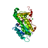
| ||||||||
|---|---|---|---|---|---|---|---|---|---|
| 1 | 
| ||||||||
| Unit cell |
|
-Components
| #1: Protein | Mass: 30864.008 Da / Num. of mol.: 1 Source method: isolated from a genetically manipulated source Source: (gene. exp.) Moraxella catarrhalis (strain BBH18) (bacteria)Strain: BBH18 / Gene: fabI, MCR_1078 / Plasmid: pET28C / Production host: References: UniProt: D5VCE0, enoyl-[acyl-carrier-protein] reductase (NADH) |
|---|---|
| #2: Chemical | ChemComp-NAD /   Mass: 663.425 Da / Num. of mol.: 1 / Source method: obtained synthetically / Formula: C21H27N7O14P2 / Feature type: SUBJECT OF INVESTIGATION / Comment: NAD*YM Mass: 663.425 Da / Num. of mol.: 1 / Source method: obtained synthetically / Formula: C21H27N7O14P2 / Feature type: SUBJECT OF INVESTIGATION / Comment: NAD*YM |
| #3: Chemical | ChemComp-CA /   Mass: 40.078 Da / Num. of mol.: 1 / Source method: obtained synthetically / Formula: Ca Mass: 40.078 Da / Num. of mol.: 1 / Source method: obtained synthetically / Formula: Ca |
| #4: Chemical | ChemComp-GOL /   Mass: 92.094 Da / Num. of mol.: 1 / Source method: obtained synthetically / Formula: C3H8O3 Mass: 92.094 Da / Num. of mol.: 1 / Source method: obtained synthetically / Formula: C3H8O3 |
| #5: Water | ChemComp-HOH /  Mass: 18.015 Da / Num. of mol.: 56 / Source method: isolated from a natural source / Formula: H2O Mass: 18.015 Da / Num. of mol.: 56 / Source method: isolated from a natural source / Formula: H2O |
| Has ligand of interest | Y |
-Experimental details
-Experiment
| Experiment | Method: X-RAY DIFFRACTION / Number of used crystals: 1 |
|---|
- Sample preparation
| Crystal | Density Matthews: 2.5 Å3/Da / Density % sol: 50.92 % / Description: Rod shaped crystals |
|---|---|
| Crystal grow | Temperature: 293 K / Method: vapor diffusion, sitting drop / pH: 7 Details: 0.2M calcium chloride, 0.1M HEPES buffer, 22% PEG 400, 10-folds of NADH cofactor |
-Data collection
| Diffraction | Mean temperature: 100 K / Serial crystal experiment: N | ||||||||||||||||||||||||||||||
|---|---|---|---|---|---|---|---|---|---|---|---|---|---|---|---|---|---|---|---|---|---|---|---|---|---|---|---|---|---|---|---|
| Diffraction source | Source: SYNCHROTRON / Site: ESRF  / Beamline: BM30A / Wavelength: 0.9677 Å / Beamline: BM30A / Wavelength: 0.9677 Å | ||||||||||||||||||||||||||||||
| Detector | Type: DECTRIS EIGER X 4M / Detector: PIXEL / Date: Mar 8, 2018 | ||||||||||||||||||||||||||||||
| Radiation | Protocol: SINGLE WAVELENGTH / Monochromatic (M) / Laue (L): M / Scattering type: x-ray | ||||||||||||||||||||||||||||||
| Radiation wavelength | Wavelength: 0.9677 Å / Relative weight: 1 | ||||||||||||||||||||||||||||||
| Reflection | Resolution: 2.12→60.7 Å / Num. obs: 17427 / % possible obs: 99.9 % / Redundancy: 11.1 % / CC1/2: 1 / Rmerge(I) obs: 0.048 / Rpim(I) all: 0.015 / Rrim(I) all: 0.051 / Net I/σ(I): 22.3 | ||||||||||||||||||||||||||||||
| Reflection shell | Diffraction-ID: 1
|
-Phasing
| Phasing | Method: molecular replacement |
|---|
- Processing
| Software |
| |||||||||||||||||||||||||||||||||||||||||||||
|---|---|---|---|---|---|---|---|---|---|---|---|---|---|---|---|---|---|---|---|---|---|---|---|---|---|---|---|---|---|---|---|---|---|---|---|---|---|---|---|---|---|---|---|---|---|---|
| Refinement | Method to determine structure: MOLECULAR REPLACEMENT Starting model: 7CPG 7cpg Resolution: 2.12→47.59 Å / Cor.coef. Fo:Fc: 0.97 / Cor.coef. Fo:Fc free: 0.946 / SU B: 7.786 / SU ML: 0.189 / SU R Cruickshank DPI: 0.2177 / Cross valid method: THROUGHOUT / σ(F): 0 / ESU R: 0.218 / ESU R Free: 0.196 / Stereochemistry target values: MAXIMUM LIKELIHOOD / Details: U VALUES : REFINED INDIVIDUALLY
| |||||||||||||||||||||||||||||||||||||||||||||
| Solvent computation | Ion probe radii: 0.8 Å / Shrinkage radii: 0.8 Å / VDW probe radii: 1.2 Å / Solvent model: MASK | |||||||||||||||||||||||||||||||||||||||||||||
| Displacement parameters | Biso max: 154.1 Å2 / Biso mean: 69.505 Å2 / Biso min: 39.97 Å2
| |||||||||||||||||||||||||||||||||||||||||||||
| Refinement step | Cycle: final / Resolution: 2.12→47.59 Å
| |||||||||||||||||||||||||||||||||||||||||||||
| Refine LS restraints |
| |||||||||||||||||||||||||||||||||||||||||||||
| LS refinement shell | Resolution: 2.121→2.176 Å / Rfactor Rfree error: 0 / Total num. of bins used: 20
|
