 Movie
Movie Controller

[English] 日本語
 Yorodumi
- PDB-7f23: Cryo-EM structure of the GTP-bound dopamine receptor 1 and mini-G... -
+ Open data

- Basic information
| Entry | Database: PDB / ID: 7f23 | |||||||||||||||||||||||||||
|---|---|---|---|---|---|---|---|---|---|---|---|---|---|---|---|---|---|---|---|---|---|---|---|---|---|---|---|---|
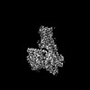
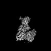
| Title | Cryo-EM structure of the GTP-bound dopamine receptor 1 and mini-Gs complex with Nb35 | |||||||||||||||||||||||||||
 Components Components |
| |||||||||||||||||||||||||||
 Keywords Keywords | MEMBRANE PROTEIN / GPCR / dopamine receptor / mini-Gs | |||||||||||||||||||||||||||
| Function / homology |  Function and homology information Function and homology informationdopamine neurotransmitter receptor activity, coupled via Gs / dopamine neurotransmitter receptor activity / cerebral cortex GABAergic interneuron migration / Dopamine receptors / regulation of dopamine uptake involved in synaptic transmission / dopamine binding / operant conditioning / phospholipase C-activating dopamine receptor signaling pathway / heterotrimeric G-protein binding / peristalsis ...dopamine neurotransmitter receptor activity, coupled via Gs / dopamine neurotransmitter receptor activity / cerebral cortex GABAergic interneuron migration / Dopamine receptors / regulation of dopamine uptake involved in synaptic transmission / dopamine binding / operant conditioning / phospholipase C-activating dopamine receptor signaling pathway / heterotrimeric G-protein binding / peristalsis / G protein-coupled receptor complex / modification of postsynaptic structure / positive regulation of neuron migration / habituation / grooming behavior / regulation of dopamine metabolic process / : / striatum development / astrocyte development / dentate gyrus development / positive regulation of potassium ion transport / sensitization / conditioned taste aversion / maternal behavior / arrestin family protein binding / non-motile cilium / adult walking behavior / G protein-coupled dopamine receptor signaling pathway / long-term synaptic depression / mating behavior / : / ciliary membrane / dopamine metabolic process / temperature homeostasis / adenylate cyclase-activating G protein-coupled bile acid receptor signaling pathway / adenylate cyclase-activating serotonin receptor signaling pathway / transmission of nerve impulse / regulation of skeletal muscle contraction / PKA activation in glucagon signalling / hair follicle placode formation / developmental growth / intracellular transport / G-protein alpha-subunit binding / G protein-coupled receptor signaling pathway, coupled to cyclic nucleotide second messenger / D1 dopamine receptor binding / vascular endothelial cell response to laminar fluid shear stress / renal water homeostasis / behavioral fear response / Hedgehog 'off' state / activation of adenylate cyclase activity / adenylate cyclase-activating adrenergic receptor signaling pathway / neuronal action potential / cellular response to acidic pH / positive regulation of synaptic transmission, glutamatergic / behavioral response to cocaine / synapse assembly / cellular response to glucagon stimulus / intracellular glucose homeostasis / presynaptic modulation of chemical synaptic transmission / positive regulation of insulin secretion involved in cellular response to glucose stimulus / adenylate cyclase activator activity / trans-Golgi network membrane / positive regulation of release of sequestered calcium ion into cytosol / response to amphetamine / negative regulation of inflammatory response to antigenic stimulus / synaptic transmission, glutamatergic / response to prostaglandin E / bone development / visual learning / platelet aggregation / cognition / GABA-ergic synapse / vasodilation / G protein-coupled receptor activity / G-protein beta/gamma-subunit complex binding / memory / positive regulation of insulin secretion / protein import into nucleus / Olfactory Signaling Pathway / Activation of the phototransduction cascade / G protein-coupled acetylcholine receptor signaling pathway / G beta:gamma signalling through PLC beta / Presynaptic function of Kainate receptors / Thromboxane signalling through TP receptor / sensory perception of smell / Activation of G protein gated Potassium channels / Inhibition of voltage gated Ca2+ channels via Gbeta/gamma subunits / G-protein activation / Glucagon signaling in metabolic regulation / Prostacyclin signalling through prostacyclin receptor / G beta:gamma signalling through CDC42 / Synthesis, secretion, and inactivation of Glucagon-like Peptide-1 (GLP-1) / G beta:gamma signalling through BTK / photoreceptor disc membrane / ADP signalling through P2Y purinoceptor 12 / Glucagon-type ligand receptors / long-term synaptic potentiation / Sensory perception of sweet, bitter, and umami (glutamate) taste / Adrenaline,noradrenaline inhibits insulin secretion / Vasopressin regulates renal water homeostasis via Aquaporins Similarity search - Function | |||||||||||||||||||||||||||
| Biological species |  Homo sapiens (human) Homo sapiens (human)synthetic construct (others) | |||||||||||||||||||||||||||
| Method | ELECTRON MICROSCOPY / single particle reconstruction / cryo EM / Resolution: 3.58 Å | |||||||||||||||||||||||||||
 Authors Authors | Xiao, T. / Zheng, S. | |||||||||||||||||||||||||||
| Funding support |  China, 1items China, 1items
| |||||||||||||||||||||||||||
 Citation Citation | Journal: Sci Adv / Year: 2022 Title: Structural insights into G protein activation by D1 dopamine receptor. Authors: Xiao Teng / Sijia Chen / Qing Wang / Zhao Chen / Xiaoying Wang / Niu Huang / Sanduo Zheng / Abstract: G protein-coupled receptors (GPCRs) comprise the largest family of membrane receptors and are the most important drug targets. An agonist-bound GPCR engages heterotrimeric G proteins and triggers the ...G protein-coupled receptors (GPCRs) comprise the largest family of membrane receptors and are the most important drug targets. An agonist-bound GPCR engages heterotrimeric G proteins and triggers the exchange of guanosine diphosphate (GDP) with guanosine triphosphate (GTP) to promote G protein activation. A complete understanding of molecular mechanisms of G protein activation has been hindered by a lack of structural information of GPCR-G protein complex in nucleotide-bound states. Here, we report the cryo-EM structures of the D1 dopamine receptor and mini-G complex in the nucleotide-free and nucleotide-bound states. These structures reveal major conformational changes in Gα such as structural rearrangements of the carboxyl- and amino-terminal α helices that account for the release of GDP and the GTP-dependent dissociation of Gα from Gβγ subunits. As validated by biochemical and cellular signaling studies, our structures shed light into the molecular basis of the entire signaling events of GPCR-mediated G protein activation. | |||||||||||||||||||||||||||
| History |
|
- Structure visualization
| Structure viewer | Molecule: MolmilJmol/JSmol |
|---|
- Downloads & links
-Download
| PDBx/mmCIF format | 7f23.cif.gz | 230.8 KB | Display | PDBx/mmCIF format |
|---|---|---|---|---|
| PDB format | pdb7f23.ent.gz | 172.4 KB | Display | PDB format |
| PDBx/mmJSON format | 7f23.json.gz | Tree view | PDBx/mmJSON format | |
| Others |  Other downloads Other downloads |
-Validation report
| Arichive directory | https://data.pdbj.org/pub/pdb/validation_reports/f2/7f23ftp://data.pdbj.org/pub/pdb/validation_reports/f2/7f23 | HTTPS FTP |
|---|
-Related structure data
| Related structure data |  31426MC 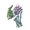 7f0tC  7f1oC 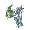 7f1zC 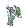 7f24C M: map data used to model this data C: citing same article ( |
|---|---|
| Similar structure data |


-Links
 PDBj
PDBj


- Assembly
| Deposited unit | 
|
|---|---|
| 1 |
|
-Components
-Guanine nucleotide-binding protein ... , 3 types, 3 molecules ABD
| #2: Protein | Mass: 28907.684 Da / Num. of mol.: 1 / Mutation: G49D, E50N, A235D, S238D, I358A, V361I Source method: isolated from a genetically manipulated source Source: (gene. exp.) Homo sapiens (human) / Gene: GNAS, GNAS1, GSP / Production host: Homo sapiens (human) / References: UniProt: P63092 |
|---|---|
| #3: Protein | Mass: 39418.086 Da / Num. of mol.: 1 Source method: isolated from a genetically manipulated source Source: (gene. exp.) Homo sapiens (human) / Gene: GNB1 / Production host:  Trichoplusia ni (cabbage looper) / References: UniProt: P62873 Trichoplusia ni (cabbage looper) / References: UniProt: P62873 |
| #5: Protein | Mass: 7845.078 Da / Num. of mol.: 1 / Mutation: C68S Source method: isolated from a genetically manipulated source Source: (gene. exp.) Homo sapiens (human) / Gene: GNG2 / Production host: Trichoplusia ni (cabbage looper) / References: UniProt: P59768 |
-Protein / Antibody , 2 types, 2 molecules FE
| #1: Protein | Mass: 52409.656 Da / Num. of mol.: 1 Source method: isolated from a genetically manipulated source Source: (gene. exp.) Homo sapiens (human) / Gene: DRD1 / Production host: Homo sapiens (human) / References: UniProt: P21728 |
|---|---|
| #4: Antibody | Mass: 17352.498 Da / Num. of mol.: 1 Source method: isolated from a genetically manipulated source Source: (gene. exp.) synthetic construct (others) / Production host:  |
-Non-polymers , 2 types, 2 molecules 

| #6: Chemical | ChemComp-LDP /  Mass: 153.178 Da / Num. of mol.: 1 / Source method: obtained synthetically / Formula: C8H11NO2 / Feature type: SUBJECT OF INVESTIGATION / Comment: medication*YM Mass: 153.178 Da / Num. of mol.: 1 / Source method: obtained synthetically / Formula: C8H11NO2 / Feature type: SUBJECT OF INVESTIGATION / Comment: medication*YM |
|---|---|
| #7: Chemical | ChemComp-GTP /  Mass: 523.180 Da / Num. of mol.: 1 / Source method: obtained synthetically / Formula: C10H16N5O14P3 / Comment: GTP, energy-carrying molecule*YM Mass: 523.180 Da / Num. of mol.: 1 / Source method: obtained synthetically / Formula: C10H16N5O14P3 / Comment: GTP, energy-carrying molecule*YM |
-Details
| Has ligand of interest | Y |
|---|---|
| Has protein modification | Y |
-Experimental details
-Experiment
| Experiment | Method: ELECTRON MICROSCOPY |
|---|---|
| EM experiment | Aggregation state: PARTICLE / 3D reconstruction method: single particle reconstruction |
- Sample preparation
| Component | Name: Cryo-EM structure of the GTP-bound dopamine receptor 1 and mini-Gs complex with Nb35 Type: COMPLEX / Entity ID: #1-#5 / Source: RECOMBINANT |
|---|---|
| Source (natural) | Organism: Homo sapiens (human) |
| Source (recombinant) | Organism: Homo sapiens (human) |
| Buffer solution | pH: 7.5 |
| Specimen | Conc.: 4 mg/ml / Embedding applied: NO / Shadowing applied: NO / Staining applied: NO / Vitrification applied: YES |
| Specimen support | Grid material: GOLD / Grid mesh size: 300 divisions/in. / Grid type: Quantifoil R1.2/1.3 |
| Vitrification | Instrument: FEI VITROBOT MARK IV / Cryogen name: ETHANE / Humidity: 100 % / Chamber temperature: 281 K |
- Electron microscopy imaging
| Experimental equipment |  Model: Titan Krios / Image courtesy: FEI Company |
|---|---|
| Microscopy | Model: FEI TITAN KRIOS |
| Electron gun | Electron source:  FIELD EMISSION GUN / Accelerating voltage: 300 kV / Illumination mode: FLOOD BEAM FIELD EMISSION GUN / Accelerating voltage: 300 kV / Illumination mode: FLOOD BEAM |
| Electron lens | Mode: BRIGHT FIELD / Nominal magnification: 64000 X |
| Specimen holder | Cryogen: NITROGEN |
| Image recording | Electron dose: 50 e/Å2 / Film or detector model: GATAN K3 BIOQUANTUM (6k x 4k) / Num. of real images: 1242 |
- Processing
| Software | Name: PHENIX / Version: 1.16_3549: / Classification: refinement | ||||||||||||||||||||||||
|---|---|---|---|---|---|---|---|---|---|---|---|---|---|---|---|---|---|---|---|---|---|---|---|---|---|
| EM software |
| ||||||||||||||||||||||||
| CTF correction | Type: PHASE FLIPPING AND AMPLITUDE CORRECTION | ||||||||||||||||||||||||
| Particle selection | Num. of particles selected: 1968728 | ||||||||||||||||||||||||
| 3D reconstruction | Resolution: 3.58 Å / Resolution method: FSC 0.143 CUT-OFF / Num. of particles: 310901 / Num. of class averages: 1 / Symmetry type: POINT | ||||||||||||||||||||||||
| Refine LS restraints |
|
