 Movie
Movie Controller


+ Open data

- Basic information
| Entry | Database: PDB / ID: 7f21 | |||||||||
|---|---|---|---|---|---|---|---|---|---|---|
| Title | L-lactate oxidase with D-lactate | |||||||||
 Components Components | L-lactate oxidase | |||||||||
 Keywords Keywords | OXIDOREDUCTASE / D-lactate / complex | |||||||||
| Function / homology | FLAVIN MONONUCLEOTIDE / LACTIC ACID Function and homology information Function and homology information | |||||||||
| Biological species |  Aerococcus viridans (bacteria) Aerococcus viridans (bacteria) | |||||||||
| Method |  X-RAY DIFFRACTION / SYNCHROTRON / MOLECULAR REPLACEMENT / Resolution: 1.38 Å X-RAY DIFFRACTION / SYNCHROTRON / MOLECULAR REPLACEMENT / Resolution: 1.38 Å | |||||||||
 Authors Authors | Morimoto, Y. / Inaka, K. | |||||||||
 Citation Citation | Journal: Biochem.Biophys.Res.Commun. / Year: 2021 Title: Dynamic interactions in the l-lactate oxidase active site facilitate substrate binding at pH4.5. Authors: Furubayashi, N. / Inaka, K. / Kamo, M. / Umena, Y. / Matsuoka, T. / Morimoto, Y. | |||||||||
| History |
|
- Structure visualization
| Structure viewer | Molecule: MolmilJmol/JSmol |
|---|
- Downloads & links
-Download
| PDBx/mmCIF format | 7f21.cif.gz | 185.9 KB | Display | PDBx/mmCIF format |
|---|---|---|---|---|
| PDB format | pdb7f21.ent.gz | 137.9 KB | Display | PDB format |
| PDBx/mmJSON format | 7f21.json.gz | Tree view | PDBx/mmJSON format | |
| Others |  Other downloads Other downloads |
-Validation report
| Arichive directory | https://data.pdbj.org/pub/pdb/validation_reports/f2/7f21ftp://data.pdbj.org/pub/pdb/validation_reports/f2/7f21 | HTTPS FTP |
|---|
-Related structure data
| Related structure data |  7f1yC  7f20C  7f22C  2du2S S: Starting model for refinement C: citing same article ( |
|---|---|
| Similar structure data |
-Links
 PDBj
PDBj- Assembly
| Deposited unit | 
| ||||||||
|---|---|---|---|---|---|---|---|---|---|
| 1 |
| ||||||||
| Unit cell |
| ||||||||
| Components on special symmetry positions |
|
-Components
| #1: Protein | Mass: 78793.289 Da / Num. of mol.: 2 Source method: isolated from a genetically manipulated source Source: (gene. exp.) Aerococcus viridans (bacteria) / Production host: Aerococcus viridans (bacteria)#2: Chemical |   Mass: 456.344 Da / Num. of mol.: 2 / Source method: obtained synthetically / Formula: C17H21N4O9P Mass: 456.344 Da / Num. of mol.: 2 / Source method: obtained synthetically / Formula: C17H21N4O9P#3: Chemical | 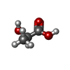  Mass: 90.078 Da / Num. of mol.: 2 / Source method: obtained synthetically / Formula: C3H6O3 / Feature type: SUBJECT OF INVESTIGATION Mass: 90.078 Da / Num. of mol.: 2 / Source method: obtained synthetically / Formula: C3H6O3 / Feature type: SUBJECT OF INVESTIGATION#4: Water | ChemComp-HOH / |  Mass: 18.015 Da / Num. of mol.: 740 / Source method: isolated from a natural source / Formula: H2O Mass: 18.015 Da / Num. of mol.: 740 / Source method: isolated from a natural source / Formula: H2OHas ligand of interest | Y | |
|---|
-Experimental details
-Experiment
| Experiment | Method: X-RAY DIFFRACTION / Number of used crystals: 1 |
|---|
- Sample preparation
| Crystal | Density Matthews: 1.29 Å3/Da / Density % sol: 4.47 % |
|---|---|
| Crystal grow | Temperature: 298 K / Method: vapor diffusion Details: 20 mg/ml in 50 mM sodium acetate (pH4.5) , 40% ethylene glycol |
-Data collection
| Diffraction | Mean temperature: 100 K / Serial crystal experiment: N |
|---|---|
| Diffraction source | Source: SYNCHROTRON / Site: SPring-8  / Beamline: BL44XU / Wavelength: 0.9 Å / Beamline: BL44XU / Wavelength: 0.9 Å |
| Detector | Type: DECTRIS EIGER X 16M / Detector: PIXEL / Date: Jul 20, 2020 |
| Radiation | Protocol: SINGLE WAVELENGTH / Monochromatic (M) / Laue (L): M / Scattering type: x-ray |
| Radiation wavelength | Wavelength: 0.9 Å / Relative weight: 1 |
| Reflection | Resolution: 1.38→46.99 Å / Num. obs: 155462 / % possible obs: 99.37 % / Redundancy: 16.6 % / CC1/2: 0.96 / Net I/σ(I): 11.65 |
| Reflection shell | Resolution: 1.38→1.429 Å / Num. unique obs: 16056 / CC1/2: 0.56 |
- Processing
| Software |
| ||||||||||||||||||||||||||||||||||||||||||||||||||||||||||||||||||||||||||||||||||||||||||||||||||||||||||||||||||||||||||||||||||||||||||||||||||||||||||||||||||||||||||||||||||||||
|---|---|---|---|---|---|---|---|---|---|---|---|---|---|---|---|---|---|---|---|---|---|---|---|---|---|---|---|---|---|---|---|---|---|---|---|---|---|---|---|---|---|---|---|---|---|---|---|---|---|---|---|---|---|---|---|---|---|---|---|---|---|---|---|---|---|---|---|---|---|---|---|---|---|---|---|---|---|---|---|---|---|---|---|---|---|---|---|---|---|---|---|---|---|---|---|---|---|---|---|---|---|---|---|---|---|---|---|---|---|---|---|---|---|---|---|---|---|---|---|---|---|---|---|---|---|---|---|---|---|---|---|---|---|---|---|---|---|---|---|---|---|---|---|---|---|---|---|---|---|---|---|---|---|---|---|---|---|---|---|---|---|---|---|---|---|---|---|---|---|---|---|---|---|---|---|---|---|---|---|---|---|---|---|
| Refinement | Method to determine structure: MOLECULAR REPLACEMENT Starting model: 2DU2 Resolution: 1.38→46.99 Å / Cor.coef. Fo:Fc: 0.965 / Cor.coef. Fo:Fc free: 0.954 / SU B: 1.598 / SU ML: 0.059 / Cross valid method: THROUGHOUT / ESU R: 0.062 / ESU R Free: 0.064 / Stereochemistry target values: MAXIMUM LIKELIHOOD / Details: HYDROGENS HAVE BEEN ADDED IN THE RIDING POSITIONS
| ||||||||||||||||||||||||||||||||||||||||||||||||||||||||||||||||||||||||||||||||||||||||||||||||||||||||||||||||||||||||||||||||||||||||||||||||||||||||||||||||||||||||||||||||||||||
| Solvent computation | Ion probe radii: 0.8 Å / Shrinkage radii: 0.8 Å / VDW probe radii: 1.2 Å / Solvent model: MASK | ||||||||||||||||||||||||||||||||||||||||||||||||||||||||||||||||||||||||||||||||||||||||||||||||||||||||||||||||||||||||||||||||||||||||||||||||||||||||||||||||||||||||||||||||||||||
| Displacement parameters | Biso mean: 23.001 Å2
| ||||||||||||||||||||||||||||||||||||||||||||||||||||||||||||||||||||||||||||||||||||||||||||||||||||||||||||||||||||||||||||||||||||||||||||||||||||||||||||||||||||||||||||||||||||||
| Refinement step | Cycle: 1 / Resolution: 1.38→46.99 Å
| ||||||||||||||||||||||||||||||||||||||||||||||||||||||||||||||||||||||||||||||||||||||||||||||||||||||||||||||||||||||||||||||||||||||||||||||||||||||||||||||||||||||||||||||||||||||
| Refine LS restraints |
|