 Movie
Movie Controller

[English] 日本語
 Yorodumi
- PDB-7f1a: Odinarchaeota tubulin (OdinTubulin) H393D mutant, in a protofilam... -
+ Open data

- Basic information
| Entry | Database: PDB / ID: 7f1a | ||||||||||||
|---|---|---|---|---|---|---|---|---|---|---|---|---|---|
| Title | Odinarchaeota tubulin (OdinTubulin) H393D mutant, in a protofilament arrangement, bound 78% GTP/22% GDP 1 K+, 1 Mg2+ | ||||||||||||
 Components Components | Tubulin-like protein | ||||||||||||
 Keywords Keywords | STRUCTURAL PROTEIN / Asgard / tubulin / GTP / filament | ||||||||||||
| Function / homology |  Function and homology information Function and homology informationmicrotubule-based process / structural constituent of cytoskeleton / microtubule / GTPase activity / GTP binding Similarity search - Function | ||||||||||||
| Biological species | Odinarchaeota archaeon | ||||||||||||
| Method |  X-RAY DIFFRACTION / SYNCHROTRON / MOLECULAR REPLACEMENT / Resolution: 1.9 Å X-RAY DIFFRACTION / SYNCHROTRON / MOLECULAR REPLACEMENT / Resolution: 1.9 Å | ||||||||||||
 Authors Authors | Robinson, R.C. / Akil, C. / Tran, L.T. | ||||||||||||
| Funding support |  Japan, Japan,  United States, 3items United States, 3items
| ||||||||||||
 Citation Citation | Journal: Sci Adv / Year: 2022 Title: Structure and dynamics of Odinarchaeota tubulin and the implications for eukaryotic microtubule evolution. Authors: Caner Akıl / Samson Ali / Linh T Tran / Jérémie Gaillard / Wenfei Li / Kenichi Hayashida / Mika Hirose / Takayuki Kato / Atsunori Oshima / Kosuke Fujishima / Laurent Blanchoin / Akihiro ...Authors: Caner Akıl / Samson Ali / Linh T Tran / Jérémie Gaillard / Wenfei Li / Kenichi Hayashida / Mika Hirose / Takayuki Kato / Atsunori Oshima / Kosuke Fujishima / Laurent Blanchoin / Akihiro Narita / Robert C Robinson /    Abstract: Tubulins are critical for the internal organization of eukaryotic cells, and understanding their emergence is an important question in eukaryogenesis. Asgard archaea are the closest known prokaryotic ...Tubulins are critical for the internal organization of eukaryotic cells, and understanding their emergence is an important question in eukaryogenesis. Asgard archaea are the closest known prokaryotic relatives to eukaryotes. Here, we elucidated the apo and nucleotide-bound x-ray structures of an Asgard tubulin from hydrothermal living Odinarchaeota (OdinTubulin). The guanosine 5'-triphosphate (GTP)-bound structure resembles a microtubule protofilament, with GTP bound between subunits, coordinating the "+" end subunit through a network of water molecules and unexpectedly by two cations. A water molecule is located suitable for GTP hydrolysis. Time course crystallography and electron microscopy revealed conformational changes on GTP hydrolysis. OdinTubulin forms tubules at high temperatures, with short curved protofilaments coiling around the tubule circumference, more similar to FtsZ, rather than running parallel to its length, as in microtubules. Thus, OdinTubulin represents an evolutionary stage intermediate between prokaryotic FtsZ and eukaryotic microtubule-forming tubulins. | ||||||||||||
| History |
|
- Structure visualization
| Structure viewer | Molecule: MolmilJmol/JSmol |
|---|
- Downloads & links
-Download
| PDBx/mmCIF format | 7f1a.cif.gz | 198.1 KB | Display | PDBx/mmCIF format |
|---|---|---|---|---|
| PDB format | pdb7f1a.ent.gz | 148.5 KB | Display | PDB format |
| PDBx/mmJSON format | 7f1a.json.gz | Tree view | PDBx/mmJSON format | |
| Others |  Other downloads Other downloads |
-Validation report
| Arichive directory | https://data.pdbj.org/pub/pdb/validation_reports/f1/7f1aftp://data.pdbj.org/pub/pdb/validation_reports/f1/7f1a | HTTPS FTP |
|---|
-Related structure data
| Related structure data | 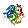 7evbSC 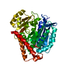 7evcC 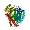 7evdC 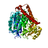 7eveC  7evgC 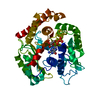 7evhC 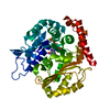 7eviC 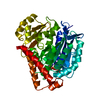 7evkC 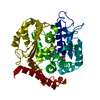 7evlC 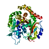 7f1bC S: Starting model for refinement C: citing same article ( |
|---|---|
| Similar structure data |


-Links
 PDBj
PDBj


- Assembly
| Deposited unit | 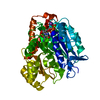
| ||||||||||||
|---|---|---|---|---|---|---|---|---|---|---|---|---|---|
| 1 |
| ||||||||||||
| Unit cell |
|
-Components
-Protein , 1 types, 1 molecules B
| #1: Protein | Mass: 47633.406 Da / Num. of mol.: 1 / Mutation: H393D Source method: isolated from a genetically manipulated source Source: (gene. exp.)  Odinarchaeota archaeon (strain LCB_4) (archaea) Odinarchaeota archaeon (strain LCB_4) (archaea)Strain: LCB_4 / Gene: cetZ, OdinLCB4_01330 / Production host:  |
|---|
-Non-polymers , 5 types, 180 molecules 


| #2: Chemical | ChemComp-GTP /  Mass: 523.180 Da / Num. of mol.: 1 / Source method: obtained synthetically / Formula: C10H16N5O14P3 / Feature type: SUBJECT OF INVESTIGATION / Comment: GTP, energy-carrying molecule*YM Mass: 523.180 Da / Num. of mol.: 1 / Source method: obtained synthetically / Formula: C10H16N5O14P3 / Feature type: SUBJECT OF INVESTIGATION / Comment: GTP, energy-carrying molecule*YM |
|---|---|
| #3: Chemical | ChemComp-GDP /  Type: RNA linking / Mass: 443.201 Da / Num. of mol.: 1 / Source method: obtained synthetically / Formula: C10H15N5O11P2 / Feature type: SUBJECT OF INVESTIGATION / Comment: GDP, energy-carrying molecule*YM Type: RNA linking / Mass: 443.201 Da / Num. of mol.: 1 / Source method: obtained synthetically / Formula: C10H15N5O11P2 / Feature type: SUBJECT OF INVESTIGATION / Comment: GDP, energy-carrying molecule*YM |
| #4: Chemical | ChemComp-MG /  Mass: 24.305 Da / Num. of mol.: 1 / Source method: obtained synthetically / Formula: Mg / Feature type: SUBJECT OF INVESTIGATION Mass: 24.305 Da / Num. of mol.: 1 / Source method: obtained synthetically / Formula: Mg / Feature type: SUBJECT OF INVESTIGATION |
| #5: Chemical | ChemComp-K /  Mass: 39.098 Da / Num. of mol.: 1 / Source method: obtained synthetically / Formula: K / Feature type: SUBJECT OF INVESTIGATION Mass: 39.098 Da / Num. of mol.: 1 / Source method: obtained synthetically / Formula: K / Feature type: SUBJECT OF INVESTIGATION |
| #6: Water | ChemComp-HOH / Mass: 18.015 Da / Num. of mol.: 176 / Source method: isolated from a natural source / Formula: H2O |
-Details
| Has ligand of interest | Y |
|---|
-Experimental details
-Experiment
| Experiment | Method: X-RAY DIFFRACTION / Number of used crystals: 1 |
|---|
- Sample preparation
| Crystal | Density Matthews: 2.02 Å3/Da / Density % sol: 39.07 % |
|---|---|
| Crystal grow | Temperature: 293 K / Method: vapor diffusion, hanging drop / pH: 9 Details: 25% PEG 1500 0.1 M MMT (1 DL-malic acid: 2 MES: 2 Tris base), pH 9.0 Soak: 10 mM GTP, 1 mM MgCl2, 100 mM KCl (1 h) |
-Data collection
| Diffraction | Mean temperature: 100 K / Serial crystal experiment: N |
|---|---|
| Diffraction source | Source: SYNCHROTRON / Site: SPring-8 / Beamline: BL41XU / Wavelength: 1 Å |
| Detector | Type: DECTRIS EIGER2 X 16M / Detector: PIXEL / Date: Jun 4, 2021 |
| Radiation | Protocol: SINGLE WAVELENGTH / Monochromatic (M) / Laue (L): M / Scattering type: x-ray |
| Radiation wavelength | Wavelength: 1 Å / Relative weight: 1 |
| Reflection | Resolution: 1.9→29.62 Å / Num. obs: 31257 / % possible obs: 100 % / Redundancy: 19.9 % / Rmerge(I) obs: 0.123 / Rpim(I) all: 0.028 / Rrim(I) all: 0.127 / Net I/σ(I): 14.9 |
| Reflection shell | Resolution: 1.9→1.94 Å / Redundancy: 20.3 % / Rmerge(I) obs: 2.337 / Mean I/σ(I) obs: 1.7 / Num. unique obs: 2053 / CC1/2: 0.75 / Rpim(I) all: 0.529 / Rrim(I) all: 2.397 / % possible all: 100 |
- Processing
| Software |
| ||||||||||||||||||||||||
|---|---|---|---|---|---|---|---|---|---|---|---|---|---|---|---|---|---|---|---|---|---|---|---|---|---|
| Refinement | Method to determine structure: MOLECULAR REPLACEMENT Starting model: 7EVB Resolution: 1.9→29.2 Å / Cross valid method: FREE R-VALUE Stereochemistry target values: GeoStd + Monomer Library + CDL v1.2
| ||||||||||||||||||||||||
| Displacement parameters | Biso mean: 38.92 Å2 | ||||||||||||||||||||||||
| Refinement step | Cycle: LAST / Resolution: 1.9→29.2 Å
| ||||||||||||||||||||||||
| Refine LS restraints |
| ||||||||||||||||||||||||
| LS refinement shell | Resolution: 1.9→1.97 Å /
|
