 Movie
Movie Controller


+ Open data

- Basic information
| Entry | Database: PDB / ID: 7f0i | ||||||
|---|---|---|---|---|---|---|---|
| Title | phosphodiesterase-9A in complex with inhibitor 4b | ||||||
 Components Components | Isoform PDE9A2 of High affinity cGMP-specific 3',5'-cyclic phosphodiesterase 9A | ||||||
 Keywords Keywords | HYDROLASE/INHIBITOR / HYDROLASE-Inhibitor complex | ||||||
| Function / homology | 3',5'-cyclic-GMP phosphodiesterase / Chem-06A / Isoform PDE9A2 of High affinity cGMP-specific 3',5'-cyclic phosphodiesterase 9A Function and homology information Function and homology information | ||||||
| Biological species |  Homo sapiens (human) Homo sapiens (human) | ||||||
| Method |  X-RAY DIFFRACTION / MOLECULAR REPLACEMENT / Resolution: 2.70000890032 Å X-RAY DIFFRACTION / MOLECULAR REPLACEMENT / Resolution: 2.70000890032 Å | ||||||
 Authors Authors | Wu, Y. / Huang, Y.Y. / Luo, H.B. | ||||||
| Funding support |  China, 1items China, 1items
| ||||||
 Citation Citation | Journal: J.Med.Chem. / Year: 2021 Title: Discovery of Potent Phosphodiesterase-9 Inhibitors for the Treatment of Hepatic Fibrosis Authors: Wu, Y. / Wang, Q. / Jiang, M.Y. / Huang, Y.Y. / Zhu, Z. / Han, C. / Tian, Y.J. / Zhang, B. / Luo, H.B. | ||||||
| History |
|
- Structure visualization
| Structure viewer | Molecule: MolmilJmol/JSmol |
|---|
- Downloads & links
-Download
| PDBx/mmCIF format | 7f0i.cif.gz | 153 KB | Display | PDBx/mmCIF format |
|---|---|---|---|---|
| PDB format | pdb7f0i.ent.gz | 114.2 KB | Display | PDB format |
| PDBx/mmJSON format | 7f0i.json.gz | Tree view | PDBx/mmJSON format | |
| Others |  Other downloads Other downloads |
-Validation report
| Arichive directory | https://data.pdbj.org/pub/pdb/validation_reports/f0/7f0iftp://data.pdbj.org/pub/pdb/validation_reports/f0/7f0i | HTTPS FTP |
|---|
-Related structure data
| Related structure data |  6a3nS S: Starting model for refinement |
|---|---|
| Similar structure data |
-Links
 PDBj
PDBj
- Assembly
| Deposited unit | 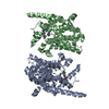
| ||||||||||||
|---|---|---|---|---|---|---|---|---|---|---|---|---|---|
| 1 | 
| ||||||||||||
| 2 | 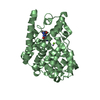
| ||||||||||||
| Unit cell |
|
-Components
| #1: Protein | Mass: 61789.078 Da / Num. of mol.: 2 Source method: isolated from a genetically manipulated source Source: (gene. exp.) Homo sapiens (human) / Gene: PDE9A / Production host:  References: UniProt: O76083-2, 3',5'-cyclic-GMP phosphodiesterase #2: Chemical |   Mass: 65.409 Da / Num. of mol.: 2 / Source method: obtained synthetically / Formula: Zn Mass: 65.409 Da / Num. of mol.: 2 / Source method: obtained synthetically / Formula: Zn#3: Chemical |   Mass: 24.305 Da / Num. of mol.: 2 / Source method: obtained synthetically / Formula: Mg Mass: 24.305 Da / Num. of mol.: 2 / Source method: obtained synthetically / Formula: Mg#4: Chemical | 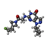  Mass: 388.439 Da / Num. of mol.: 2 / Source method: obtained synthetically / Formula: C19H25FN6O2 / Feature type: SUBJECT OF INVESTIGATION Mass: 388.439 Da / Num. of mol.: 2 / Source method: obtained synthetically / Formula: C19H25FN6O2 / Feature type: SUBJECT OF INVESTIGATION#5: Water | ChemComp-HOH / |  Mass: 18.015 Da / Num. of mol.: 116 / Source method: isolated from a natural source / Formula: H2O Mass: 18.015 Da / Num. of mol.: 116 / Source method: isolated from a natural source / Formula: H2OHas ligand of interest | Y | Sequence details | THIS SEQUENCE CORRESPOND | |
|---|
-Experimental details
-Experiment
| Experiment | Method: X-RAY DIFFRACTION / Number of used crystals: 1 |
|---|
- Sample preparation
| Crystal | Density Matthews: 2.91 Å3/Da / Density % sol: 57.76 % |
|---|---|
| Crystal grow | Temperature: 277 K / Method: vapor diffusion, hanging drop / Details: 3.0M Na formate, 0.1M HEPES (pH 7.5), 5% xylitol |
-Data collection
| Diffraction | Mean temperature: 100 K / Serial crystal experiment: N |
|---|---|
| Diffraction source | Source: ROTATING ANODE / Type: RIGAKU / Wavelength: 1.54056 Å |
| Detector | Type: RIGAKU HyPix-3000 / Detector: PIXEL / Date: Apr 12, 2021 |
| Radiation | Protocol: SINGLE WAVELENGTH / Monochromatic (M) / Laue (L): M / Scattering type: x-ray |
| Radiation wavelength | Wavelength: 1.54056 Å / Relative weight: 1 |
| Reflection | Resolution: 2.7→23.5321 Å / Num. obs: 40355 / % possible obs: 98.4 % / Redundancy: 8.9 % / Biso Wilson estimate: 34.5489656212 Å2 / Rmerge(I) obs: 0.14 / Net I/σ(I): 21.4 |
| Reflection shell | Resolution: 2.7→2.8 Å / Redundancy: 9.7 % / Rmerge(I) obs: 0.45 / Mean I/σ(I) obs: 5 / Num. unique obs: 4030 / % possible all: 100 |
- Processing
| Software |
| |||||||||||||||||||||||||||||||||||||||||||||||||||||||||||||||||||||||||||||||||||||||||||||||||||||||||
|---|---|---|---|---|---|---|---|---|---|---|---|---|---|---|---|---|---|---|---|---|---|---|---|---|---|---|---|---|---|---|---|---|---|---|---|---|---|---|---|---|---|---|---|---|---|---|---|---|---|---|---|---|---|---|---|---|---|---|---|---|---|---|---|---|---|---|---|---|---|---|---|---|---|---|---|---|---|---|---|---|---|---|---|---|---|---|---|---|---|---|---|---|---|---|---|---|---|---|---|---|---|---|---|---|---|---|
| Refinement | Method to determine structure: MOLECULAR REPLACEMENT Starting model: 6A3N Resolution: 2.70000890032→23.5320777477 Å / SU ML: 0.504562992188 / Cross valid method: NONE / σ(F): 2.02616478662 / Phase error: 32.4962839839 Stereochemistry target values: GeoStd + Monomer Library + CDL v1.2
| |||||||||||||||||||||||||||||||||||||||||||||||||||||||||||||||||||||||||||||||||||||||||||||||||||||||||
| Solvent computation | Shrinkage radii: 0.9 Å / VDW probe radii: 1.11 Å / Solvent model: FLAT BULK SOLVENT MODEL | |||||||||||||||||||||||||||||||||||||||||||||||||||||||||||||||||||||||||||||||||||||||||||||||||||||||||
| Displacement parameters | Biso mean: 30.0805812221 Å2 | |||||||||||||||||||||||||||||||||||||||||||||||||||||||||||||||||||||||||||||||||||||||||||||||||||||||||
| Refinement step | Cycle: LAST / Resolution: 2.70000890032→23.5320777477 Å
| |||||||||||||||||||||||||||||||||||||||||||||||||||||||||||||||||||||||||||||||||||||||||||||||||||||||||
| Refine LS restraints |
| |||||||||||||||||||||||||||||||||||||||||||||||||||||||||||||||||||||||||||||||||||||||||||||||||||||||||
| LS refinement shell |
|
