 Movie
Movie Controller


+ Open data

- Basic information
| Entry | Database: PDB / ID: 7ey3 | ||||||
|---|---|---|---|---|---|---|---|
| Title | Double cysteine mutations in T1 lipase | ||||||
 Components Components | Thermostable lipase | ||||||
 Keywords Keywords | HYDROLASE / disulphide bond / cysteine / lipase / site-directed mutagenesis / STRUCTURAL PROTEIN | ||||||
| Function / homology |  Function and homology information Function and homology informationtriacylglycerol lipase / triacylglycerol lipase activity / lipid catabolic process / extracellular region / metal ion binding Similarity search - Function | ||||||
| Biological species |  Geobacillus zalihae (bacteria) Geobacillus zalihae (bacteria) | ||||||
| Method |  X-RAY DIFFRACTION / MOLECULAR REPLACEMENT / Resolution: 2.04 Å X-RAY DIFFRACTION / MOLECULAR REPLACEMENT / Resolution: 2.04 Å | ||||||
 Authors Authors | Hamdan, S.H. / Leow, T.C. / Yahaya, N.M. / Ali, M.S.M. / Jonet, M.A. / Mohamad Aris, S.N.A. / Maiangwa, J. | ||||||
 Citation Citation | Journal: Appl.Microbiol.Biotechnol. / Year: 2023 Title: Knotting terminal ends of mutant T1 lipase with disulfide bond improved structure rigidity and stability. Authors: Hamdan, S.H. / Maiangwa, J. / Nezhad, N.G. / Ali, M.S.M. / Normi, Y.M. / Shariff, F.M. / Rahman, R.N.Z.R.A. / Leow, T.C. | ||||||
| History |
|
- Structure visualization
| Structure viewer | Molecule: MolmilJmol/JSmol |
|---|
- Downloads & links
-Download
| PDBx/mmCIF format | 7ey3.cif.gz | 208.4 KB | Display | PDBx/mmCIF format |
|---|---|---|---|---|
| PDB format | pdb7ey3.ent.gz | 133.3 KB | Display | PDB format |
| PDBx/mmJSON format | 7ey3.json.gz | Tree view | PDBx/mmJSON format | |
| Others |  Other downloads Other downloads |
-Validation report
| Summary document | 7ey3_validation.pdf.gz | 3.2 MB | Display | wwPDB validaton report |
|---|---|---|---|---|
| Full document | 7ey3_full_validation.pdf.gz | 3.2 MB | Display | |
| Data in XML | 7ey3_validation.xml.gz | 30.1 KB | Display | |
| Data in CIF | 7ey3_validation.cif.gz | 43.7 KB | Display | |
| Arichive directory | https://data.pdbj.org/pub/pdb/validation_reports/ey/7ey3ftp://data.pdbj.org/pub/pdb/validation_reports/ey/7ey3 | HTTPS FTP |
-Related structure data
| Related structure data |  2dsnS S: Starting model for refinement |
|---|---|
| Similar structure data |
-Links
 PDBj
PDBj- Assembly
| Deposited unit | 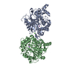
| ||||||||||||
|---|---|---|---|---|---|---|---|---|---|---|---|---|---|
| 1 | 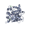
| ||||||||||||
| 2 | 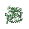
| ||||||||||||
| Unit cell |
| ||||||||||||
| Components on special symmetry positions |
|
-Components
-Protein , 1 types, 2 molecules AB
| #1: Protein | Mass: 43237.191 Da / Num. of mol.: 2 / Mutation: S2C, A384C Source method: isolated from a genetically manipulated source Source: (gene. exp.) Geobacillus zalihae (bacteria) / Production host: |
|---|
-Non-polymers , 5 types, 354 molecules 


| #2: Chemical |  Mass: 65.409 Da / Num. of mol.: 2 / Source method: obtained synthetically / Formula: Zn / Feature type: SUBJECT OF INVESTIGATION Mass: 65.409 Da / Num. of mol.: 2 / Source method: obtained synthetically / Formula: Zn / Feature type: SUBJECT OF INVESTIGATION#3: Chemical |  Mass: 40.078 Da / Num. of mol.: 2 / Source method: obtained synthetically / Formula: Ca / Feature type: SUBJECT OF INVESTIGATION Mass: 40.078 Da / Num. of mol.: 2 / Source method: obtained synthetically / Formula: Ca / Feature type: SUBJECT OF INVESTIGATION#4: Chemical |  Mass: 22.990 Da / Num. of mol.: 2 / Source method: isolated from a natural source / Formula: Na / Feature type: SUBJECT OF INVESTIGATION Mass: 22.990 Da / Num. of mol.: 2 / Source method: isolated from a natural source / Formula: Na / Feature type: SUBJECT OF INVESTIGATION#5: Chemical |  Mass: 35.453 Da / Num. of mol.: 2 / Source method: isolated from a natural source / Formula: Cl / Feature type: SUBJECT OF INVESTIGATION Mass: 35.453 Da / Num. of mol.: 2 / Source method: isolated from a natural source / Formula: Cl / Feature type: SUBJECT OF INVESTIGATION#6: Water | ChemComp-HOH / | Mass: 18.015 Da / Num. of mol.: 346 / Source method: isolated from a natural source / Formula: H2O |
|---|
-Details
| Has ligand of interest | Y |
|---|---|
| Has protein modification | Y |
-Experimental details
-Experiment
| Experiment | Method: X-RAY DIFFRACTION / Number of used crystals: 1 |
|---|
- Sample preparation
| Crystal | Density Matthews: 2.77 Å3/Da / Density % sol: 55.52 % |
|---|---|
| Crystal grow | Temperature: 293.15 K / Method: vapor diffusion, sitting drop Details: 0.5M Sodium chloride, 0.1M Sodium citrate dihydrate pH 5.6, 2 % Ethylene imine polymer |
-Data collection
| Diffraction | Mean temperature: 293.15 K / Serial crystal experiment: N |
|---|---|
| Diffraction source | Source: ROTATING ANODE / Type: RIGAKU MICROMAX-007 / Wavelength: 1.54178 Å |
| Detector | Type: RIGAKU / Detector: CCD / Date: Sep 17, 2020 |
| Radiation | Protocol: SINGLE WAVELENGTH / Monochromatic (M) / Laue (L): M / Scattering type: x-ray |
| Radiation wavelength | Wavelength: 1.54178 Å / Relative weight: 1 |
| Reflection | Resolution: 2.04→40 Å / Num. obs: 59606 / % possible obs: 99.1 % / Redundancy: 3.2 % / Biso Wilson estimate: 18.39 Å2 / CC1/2: 0.861 / Net I/σ(I): 16 |
| Reflection shell | Resolution: 2.04→2.08 Å / Num. unique obs: 107633 / CC1/2: 0.886 |
- Processing
| Software |
| |||||||||||||||||||||||||||||||||||||||||||||||||||||||||||||||||||||||||||||||||||||||||||||||||||||||||
|---|---|---|---|---|---|---|---|---|---|---|---|---|---|---|---|---|---|---|---|---|---|---|---|---|---|---|---|---|---|---|---|---|---|---|---|---|---|---|---|---|---|---|---|---|---|---|---|---|---|---|---|---|---|---|---|---|---|---|---|---|---|---|---|---|---|---|---|---|---|---|---|---|---|---|---|---|---|---|---|---|---|---|---|---|---|---|---|---|---|---|---|---|---|---|---|---|---|---|---|---|---|---|---|---|---|---|
| Refinement | Method to determine structure: MOLECULAR REPLACEMENT Starting model: 2dsn Resolution: 2.04→33.46 Å / SU ML: 0.2283 / Cross valid method: FREE R-VALUE / σ(F): 1.34 / Phase error: 22.3094 Stereochemistry target values: GeoStd + Monomer Library + CDL v1.2
| |||||||||||||||||||||||||||||||||||||||||||||||||||||||||||||||||||||||||||||||||||||||||||||||||||||||||
| Solvent computation | Shrinkage radii: 0.9 Å / VDW probe radii: 1.11 Å / Solvent model: FLAT BULK SOLVENT MODEL | |||||||||||||||||||||||||||||||||||||||||||||||||||||||||||||||||||||||||||||||||||||||||||||||||||||||||
| Displacement parameters | Biso mean: 19.92 Å2 | |||||||||||||||||||||||||||||||||||||||||||||||||||||||||||||||||||||||||||||||||||||||||||||||||||||||||
| Refinement step | Cycle: LAST / Resolution: 2.04→33.46 Å
| |||||||||||||||||||||||||||||||||||||||||||||||||||||||||||||||||||||||||||||||||||||||||||||||||||||||||
| Refine LS restraints |
| |||||||||||||||||||||||||||||||||||||||||||||||||||||||||||||||||||||||||||||||||||||||||||||||||||||||||
| LS refinement shell |
|
