 Movie
Movie Controller

[English] 日本語
 Yorodumi
- PDB-7exu: GH127 beta-L-arabinofuranosidase HypBA1 E322Q mutant complexed wi... -
+ Open data

- Basic information
| Entry | Database: PDB / ID: 7exu | ||||||||||||
|---|---|---|---|---|---|---|---|---|---|---|---|---|---|
| Title | GH127 beta-L-arabinofuranosidase HypBA1 E322Q mutant complexed with p-nitrophenyl beta-L-arabinofuranoside | ||||||||||||
 Components Components | Non-reducing end beta-L-arabinofuranosidase | ||||||||||||
 Keywords Keywords | HYDROLASE / (alpha/alpha)6 barrel / glycoside hydrolase family 127 | ||||||||||||
| Function / homology |  Function and homology information Function and homology informationnon-reducing end beta-L-arabinofuranosidase / beta-L-arabinofuranosidase activity / polysaccharide catabolic process / metal ion binding Similarity search - Function | ||||||||||||
| Biological species |  Bifidobacterium longum subsp. longum (bacteria) Bifidobacterium longum subsp. longum (bacteria) | ||||||||||||
| Method |  X-RAY DIFFRACTION / SYNCHROTRON / FOURIER SYNTHESIS / Resolution: 2.3 Å X-RAY DIFFRACTION / SYNCHROTRON / FOURIER SYNTHESIS / Resolution: 2.3 Å | ||||||||||||
 Authors Authors | Maruyama, S. / Arakawa, T. / Yamada, C. / Fujita, K. / Fushinobu, S. | ||||||||||||
| Funding support |  Japan, 3items Japan, 3items
| ||||||||||||
 Citation Citation | Journal: Glycobiology / Year: 2022 Title: Substrate complex structure, active site labeling and catalytic role of the zinc ion in cysteine glycosidase. Authors: Maruyama, S. / Sawano, K. / Amaki, S. / Suzuki, T. / Narita, S. / Kimura, K. / Arakawa, T. / Yamada, C. / Ito, Y. / Dohmae, N. / Fujita, K. / Ishiwata, A. / Fushinobu, S. #1: Journal: Biochem. Biophys. Res. Commun. / Year: 2014Title: Crystal structure of glycoside hydrolase family 127 beta-L-arabinofuranosidase from Bifidobacterium longum. Authors: Ito, T. / Saikawa, K. / Kim, S. / Fujita, K. / Ishiwata, A. / Kaeothip, S. / Arakawa, T. / Wakagi, T. / Beckham, G.T. / Ito, Y. / Fushinobu, S. | ||||||||||||
| History |
|
- Structure visualization
| Structure viewer | Molecule: MolmilJmol/JSmol |
|---|
- Downloads & links
-Download
| PDBx/mmCIF format | 7exu.cif.gz | 141.6 KB | Display | PDBx/mmCIF format |
|---|---|---|---|---|
| PDB format | pdb7exu.ent.gz | 108.7 KB | Display | PDB format |
| PDBx/mmJSON format | 7exu.json.gz | Tree view | PDBx/mmJSON format | |
| Others |  Other downloads Other downloads |
-Validation report
| Arichive directory | https://data.pdbj.org/pub/pdb/validation_reports/ex/7exuftp://data.pdbj.org/pub/pdb/validation_reports/ex/7exu | HTTPS FTP |
|---|
-Related structure data
| Related structure data |  7exvC  7exwC  3wkxS S: Starting model for refinement C: citing same article ( |
|---|---|
| Similar structure data |


-Links
 PDBj
PDBj
- Assembly
| Deposited unit | 
| ||||||||
|---|---|---|---|---|---|---|---|---|---|
| 1 | 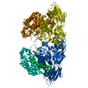
| ||||||||
| Unit cell |
|
-Components
| #1: Protein | Mass: 74456.922 Da / Num. of mol.: 1 / Mutation: E322Q Source method: isolated from a genetically manipulated source Source: (gene. exp.) Bifidobacterium longum subsp. longum (strain ATCC 15707 / DSM 20219 / JCM 1217 / NCTC 11818 / E194b) (bacteria)Strain: ATCC 15707 / DSM 20219 / JCM 1217 / NCTC 11818 / E194b Gene: hypBA1, BLLJ_0211 / Plasmid: pET23 / Production host: References: UniProt: E8MGH8, non-reducing end beta-L-arabinofuranosidase |
|---|---|
| #2: Chemical | ChemComp-ZN /   Mass: 65.409 Da / Num. of mol.: 1 / Source method: obtained synthetically / Formula: Zn / Feature type: SUBJECT OF INVESTIGATION Mass: 65.409 Da / Num. of mol.: 1 / Source method: obtained synthetically / Formula: Zn / Feature type: SUBJECT OF INVESTIGATION |
| #3: Sugar | ChemComp-07Y / (  Type: saccharide / Mass: 271.223 Da / Num. of mol.: 1 / Source method: obtained synthetically / Formula: C11H13NO7 / Feature type: SUBJECT OF INVESTIGATION Type: saccharide / Mass: 271.223 Da / Num. of mol.: 1 / Source method: obtained synthetically / Formula: C11H13NO7 / Feature type: SUBJECT OF INVESTIGATION |
| #4: Water | ChemComp-HOH /  Mass: 18.015 Da / Num. of mol.: 25 / Source method: isolated from a natural source / Formula: H2O Mass: 18.015 Da / Num. of mol.: 25 / Source method: isolated from a natural source / Formula: H2O |
| Has ligand of interest | Y |
-Experimental details
-Experiment
| Experiment | Method: X-RAY DIFFRACTION / Number of used crystals: 1 |
|---|
- Sample preparation
| Crystal | Density Matthews: 3 Å3/Da / Density % sol: 59.05 % |
|---|---|
| Crystal grow | Temperature: 293.15 K / Method: vapor diffusion, hanging drop / pH: 6.5 Details: 0.7 M sodium citrate, 0.1 M MES-NaOH (pH 6.5), 10 mM DTT |
-Data collection
| Diffraction | Mean temperature: 100 K / Serial crystal experiment: N |
|---|---|
| Diffraction source | Source: SYNCHROTRON / Site: Photon Factory / Beamline: AR-NE3A / Wavelength: 1.1 Å |
| Detector | Type: ADSC QUANTUM 270 / Detector: CCD / Date: May 27, 2015 |
| Radiation | Monochromator: Numerical link type Si(111) double crystal / Protocol: SINGLE WAVELENGTH / Monochromatic (M) / Laue (L): M / Scattering type: x-ray |
| Radiation wavelength | Wavelength: 1.1 Å / Relative weight: 1 |
| Reflection | Resolution: 2.3→46.31 Å / Num. obs: 41037 / % possible obs: 100 % / Redundancy: 10.9 % / Biso Wilson estimate: 49.46 Å2 / CC1/2: 0.999 / Rmerge(I) obs: 0.113 / Rpim(I) all: 0.036 / Net I/σ(I): 14.5 |
| Reflection shell | Resolution: 2.3→2.38 Å / Redundancy: 11.1 % / Rmerge(I) obs: 3.663 / Mean I/σ(I) obs: 1 / Num. unique obs: 3910 / CC1/2: 0.728 / Rpim(I) all: 1.144 / % possible all: 100 |
- Processing
| Software |
| ||||||||||||||||||||||||||||||||||||||||||||||||||||||||||||
|---|---|---|---|---|---|---|---|---|---|---|---|---|---|---|---|---|---|---|---|---|---|---|---|---|---|---|---|---|---|---|---|---|---|---|---|---|---|---|---|---|---|---|---|---|---|---|---|---|---|---|---|---|---|---|---|---|---|---|---|---|---|
| Refinement | Method to determine structure: FOURIER SYNTHESIS Starting model: 3WKX Resolution: 2.3→46.31 Å / Cor.coef. Fo:Fc: 0.942 / Cor.coef. Fo:Fc free: 0.92 / SU B: 15.275 / SU ML: 0.392 / Cross valid method: THROUGHOUT / σ(F): 0 / ESU R: 0.313 / ESU R Free: 0.247 / Stereochemistry target values: MAXIMUM LIKELIHOOD Details: HYDROGENS HAVE BEEN ADDED IN THE RIDING POSITIONS U VALUES : REFINED INDIVIDUALLY
| ||||||||||||||||||||||||||||||||||||||||||||||||||||||||||||
| Solvent computation | Ion probe radii: 0.8 Å / Shrinkage radii: 0.8 Å / VDW probe radii: 1.2 Å / Solvent model: BABINET MODEL WITH MASK | ||||||||||||||||||||||||||||||||||||||||||||||||||||||||||||
| Displacement parameters | Biso max: 149.13 Å2 / Biso mean: 68.703 Å2 / Biso min: 44.98 Å2
| ||||||||||||||||||||||||||||||||||||||||||||||||||||||||||||
| Refinement step | Cycle: final / Resolution: 2.3→46.31 Å
| ||||||||||||||||||||||||||||||||||||||||||||||||||||||||||||
| Refine LS restraints |
| ||||||||||||||||||||||||||||||||||||||||||||||||||||||||||||
| LS refinement shell | Resolution: 2.3→2.36 Å / Rfactor Rfree error: 0 / Total num. of bins used: 20
|
