 Movie
Movie Controller

[English] 日本語
 Yorodumi
- PDB-7exj: Crystal structure of alkaline alpha-galctosidase D383A mutant fro... -
+ Open data

- Basic information
| Entry | Database: PDB / ID: 7exj | ||||||
|---|---|---|---|---|---|---|---|
| Title | Crystal structure of alkaline alpha-galctosidase D383A mutant from Arabidopsis thaliana complexed with Raffinose | ||||||
 Components Components | Probable galactinol--sucrose galactosyltransferase 6 | ||||||
 Keywords Keywords | TRANSFERASE / Alkaline alpha-galactosidase / HYDROLASE | ||||||
| Function / homology |  Function and homology information Function and homology informationgalactinol-sucrose galactosyltransferase / galactinol-sucrose galactosyltransferase activity / plasmodesma / response to cold / chloroplast / response to oxidative stress / cellular response to hypoxia / mRNA binding / cytosol Similarity search - Function | ||||||
| Biological species |  | ||||||
| Method |  X-RAY DIFFRACTION / SYNCHROTRON / MOLECULAR REPLACEMENT / molecular replacement / Resolution: 2.47 Å X-RAY DIFFRACTION / SYNCHROTRON / MOLECULAR REPLACEMENT / molecular replacement / Resolution: 2.47 Å | ||||||
 Authors Authors | Chuankhayan, P. / Guan, H.H. / Lin, C.C. / Chen, N.C. / Huang, Y.C. / Yoshimura, M. / Nakagawa, A. / Lee, R.H. / Chen, C.J. | ||||||
 Citation Citation | Journal: Acta Crystallogr D Struct Biol / Year: 2023 Title: Structural insight into the hydrolase and synthase activities of an alkaline alpha-galactosidase from Arabidopsis from complexes with substrate/product. Authors: Chuankhayan, P. / Lee, R.H. / Guan, H.H. / Lin, C.C. / Chen, N.C. / Huang, Y.C. / Yoshimura, M. / Nakagawa, A. / Chen, C.J. | ||||||
| History |
|
- Structure visualization
| Structure viewer | Molecule: MolmilJmol/JSmol |
|---|
- Downloads & links
-Download
| PDBx/mmCIF format | 7exj.cif.gz | 291.2 KB | Display | PDBx/mmCIF format |
|---|---|---|---|---|
| PDB format | pdb7exj.ent.gz | 231.9 KB | Display | PDB format |
| PDBx/mmJSON format | 7exj.json.gz | Tree view | PDBx/mmJSON format | |
| Others |  Other downloads Other downloads |
-Validation report
| Arichive directory | https://data.pdbj.org/pub/pdb/validation_reports/ex/7exjftp://data.pdbj.org/pub/pdb/validation_reports/ex/7exj | HTTPS FTP |
|---|
-Related structure data
| Related structure data |  7exfC  7exgSC  7exhC  7exqC  7exrC S: Starting model for refinement C: citing same article ( |
|---|---|
| Similar structure data |
-Links
 PDBj
PDBj- Assembly
| Deposited unit | 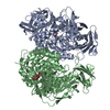
| ||||||||
|---|---|---|---|---|---|---|---|---|---|
| 1 | 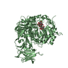
| ||||||||
| 2 | 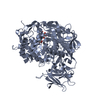
| ||||||||
| Unit cell |
|
-Components
| #1: Protein | Mass: 83204.406 Da / Num. of mol.: 2 / Mutation: D383A Source method: isolated from a genetically manipulated source Source: (gene. exp.)  References: UniProt: Q8RX87, galactinol-sucrose galactosyltransferase #2: Polysaccharide | 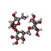  Source method: isolated from a genetically manipulated source Details: oligosaccharide with reducing-end-to-reducing-end glycosidic bond References: raffinose #3: Water | ChemComp-HOH / |  Mass: 18.015 Da / Num. of mol.: 16 / Source method: isolated from a natural source / Formula: H2O Mass: 18.015 Da / Num. of mol.: 16 / Source method: isolated from a natural source / Formula: H2OHas ligand of interest | Y | |
|---|
-Experimental details
-Experiment
| Experiment | Method: X-RAY DIFFRACTION / Number of used crystals: 1 |
|---|
- Sample preparation
| Crystal | Density Matthews: 2.61 Å3/Da / Density % sol: 52.8 % |
|---|---|
| Crystal grow | Temperature: 291.5 K / Method: microbatch / pH: 7.8 / Details: Tris, PEG 2000, PGA |
-Data collection
| Diffraction | Mean temperature: 110 K / Serial crystal experiment: N | ||||||||||||||||||||||||||||||||||||||||||||||||||||||||||||||||||||||||||||||||||||||||||||||||||||
|---|---|---|---|---|---|---|---|---|---|---|---|---|---|---|---|---|---|---|---|---|---|---|---|---|---|---|---|---|---|---|---|---|---|---|---|---|---|---|---|---|---|---|---|---|---|---|---|---|---|---|---|---|---|---|---|---|---|---|---|---|---|---|---|---|---|---|---|---|---|---|---|---|---|---|---|---|---|---|---|---|---|---|---|---|---|---|---|---|---|---|---|---|---|---|---|---|---|---|---|---|---|
| Diffraction source | Source: SYNCHROTRON / Site: SPring-8  / Beamline: BL44XU / Wavelength: 0.9 Å / Beamline: BL44XU / Wavelength: 0.9 Å | ||||||||||||||||||||||||||||||||||||||||||||||||||||||||||||||||||||||||||||||||||||||||||||||||||||
| Detector | Type: DECTRIS EIGER X 16M / Detector: PIXEL / Date: Apr 22, 2018 | ||||||||||||||||||||||||||||||||||||||||||||||||||||||||||||||||||||||||||||||||||||||||||||||||||||
| Radiation | Protocol: SINGLE WAVELENGTH / Monochromatic (M) / Laue (L): M / Scattering type: x-ray | ||||||||||||||||||||||||||||||||||||||||||||||||||||||||||||||||||||||||||||||||||||||||||||||||||||
| Radiation wavelength | Wavelength: 0.9 Å / Relative weight: 1 | ||||||||||||||||||||||||||||||||||||||||||||||||||||||||||||||||||||||||||||||||||||||||||||||||||||
| Reflection | Resolution: 2.47→30 Å / Num. obs: 118862 / % possible obs: 98.5 % / Redundancy: 3.116 % / Biso Wilson estimate: 67.722 Å2 / CC1/2: 0.997 / Rmerge(I) obs: 0.061 / Rrim(I) all: 0.074 / Χ2: 1.254 / Net I/σ(I): 10.56 / Num. measured all: 370410 | ||||||||||||||||||||||||||||||||||||||||||||||||||||||||||||||||||||||||||||||||||||||||||||||||||||
| Reflection shell | Diffraction-ID: 1
|
-Phasing
| Phasing | Method: molecular replacement |
|---|
- Processing
| Software |
| ||||||||||||||||||||||||||||||||||||||||||||||||||||||||||||
|---|---|---|---|---|---|---|---|---|---|---|---|---|---|---|---|---|---|---|---|---|---|---|---|---|---|---|---|---|---|---|---|---|---|---|---|---|---|---|---|---|---|---|---|---|---|---|---|---|---|---|---|---|---|---|---|---|---|---|---|---|---|
| Refinement | Method to determine structure: MOLECULAR REPLACEMENT Starting model: 7EXG Resolution: 2.47→30 Å / Cor.coef. Fo:Fc: 0.962 / Cor.coef. Fo:Fc free: 0.935 / SU B: 12.538 / SU ML: 0.264 / Cross valid method: THROUGHOUT / σ(F): 0 / ESU R: 0.427 / ESU R Free: 0.273 / Stereochemistry target values: MAXIMUM LIKELIHOOD Details: HYDROGENS HAVE BEEN ADDED IN THE RIDING POSITIONS U VALUES : REFINED INDIVIDUALLY
| ||||||||||||||||||||||||||||||||||||||||||||||||||||||||||||
| Solvent computation | Ion probe radii: 0.8 Å / Shrinkage radii: 0.8 Å / VDW probe radii: 1.2 Å / Solvent model: MASK | ||||||||||||||||||||||||||||||||||||||||||||||||||||||||||||
| Displacement parameters | Biso max: 167.37 Å2 / Biso mean: 71.143 Å2 / Biso min: 35.44 Å2
| ||||||||||||||||||||||||||||||||||||||||||||||||||||||||||||
| Refinement step | Cycle: final / Resolution: 2.47→30 Å
| ||||||||||||||||||||||||||||||||||||||||||||||||||||||||||||
| Refine LS restraints |
| ||||||||||||||||||||||||||||||||||||||||||||||||||||||||||||
| LS refinement shell | Resolution: 2.47→2.534 Å / Rfactor Rfree error: 0 / Total num. of bins used: 20
|
