 Movie
Movie Controller

[English] 日本語
 Yorodumi
- PDB-7euq: Crystal structure of C86H-Y124N-G126H-H196S mutant of N(omega)-hy... -
+ Open data

- Basic information
| Entry | Database: PDB / ID: 7euq | ||||||
|---|---|---|---|---|---|---|---|
| Title | Crystal structure of C86H-Y124N-G126H-H196S mutant of N(omega)-hydroxy-L-arginine hydrolase | ||||||
 Components Components | N(omega)-hydroxy-L-arginine amidinohydrolase | ||||||
 Keywords Keywords | ANTIBIOTIC / hydrolase | ||||||
| Function / homology |  Function and homology information Function and homology informationNomega-hydroxy-L-arginine amidinohydrolase / hydrolase activity, acting on carbon-nitrogen (but not peptide) bonds, in linear amidines / arginase activity / antibiotic biosynthetic process / manganese ion binding / cytosol Similarity search - Function | ||||||
| Biological species |  Streptomyces lavendulae (bacteria) Streptomyces lavendulae (bacteria) | ||||||
| Method |  X-RAY DIFFRACTION / SYNCHROTRON / MOLECULAR REPLACEMENT / Resolution: 1.8 Å X-RAY DIFFRACTION / SYNCHROTRON / MOLECULAR REPLACEMENT / Resolution: 1.8 Å | ||||||
 Authors Authors | Oda, K. / Matoba, Y. | ||||||
 Citation Citation | Journal: Protein Sci. / Year: 2022 Title: Catalytic mechanism of DcsB: Arginase framework used for hydrolyzing its inhibitor. Authors: Oda, K. / Sakaguchi, T. / Matoba, Y. | ||||||
| History |
|
- Structure visualization
| Structure viewer | Molecule: MolmilJmol/JSmol |
|---|
- Downloads & links
-Download
| PDBx/mmCIF format | 7euq.cif.gz | 121.6 KB | Display | PDBx/mmCIF format |
|---|---|---|---|---|
| PDB format | pdb7euq.ent.gz | 91.5 KB | Display | PDB format |
| PDBx/mmJSON format | 7euq.json.gz | Tree view | PDBx/mmJSON format | |
| Others |  Other downloads Other downloads |
-Validation report
| Arichive directory | https://data.pdbj.org/pub/pdb/validation_reports/eu/7euqftp://data.pdbj.org/pub/pdb/validation_reports/eu/7euq | HTTPS FTP |
|---|
-Related structure data
| Related structure data |  7eukC  7eulSC  7eunC S: Starting model for refinement C: citing same article ( |
|---|---|
| Similar structure data |
-Links
 PDBj
PDBj- Assembly
| Deposited unit | 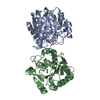
| ||||||||
|---|---|---|---|---|---|---|---|---|---|
| 1 | 
| ||||||||
| 2 | 
| ||||||||
| Unit cell |
|
-Components
| #1: Protein | Mass: 30032.662 Da / Num. of mol.: 2 / Fragment: N(omega)-hydroxy-L-arginine amidinohydrolase / Mutation: C86H, Y124N, G126H, H196S Source method: isolated from a genetically manipulated source Source: (gene. exp.) Streptomyces lavendulae (bacteria) / Gene: dcsB / Plasmid: pET21 / Production host: References: UniProt: D2Z025, Nomega-hydroxy-L-arginine amidinohydrolase #2: Chemical | ChemComp-MN /   Mass: 54.938 Da / Num. of mol.: 4 / Source method: obtained synthetically / Formula: Mn / Feature type: SUBJECT OF INVESTIGATION Mass: 54.938 Da / Num. of mol.: 4 / Source method: obtained synthetically / Formula: Mn / Feature type: SUBJECT OF INVESTIGATION#3: Chemical | ChemComp-MG /   Mass: 24.305 Da / Num. of mol.: 4 / Source method: obtained synthetically / Formula: Mg Mass: 24.305 Da / Num. of mol.: 4 / Source method: obtained synthetically / Formula: Mg#4: Water | ChemComp-HOH / |  Mass: 18.015 Da / Num. of mol.: 244 / Source method: isolated from a natural source / Formula: H2O Mass: 18.015 Da / Num. of mol.: 244 / Source method: isolated from a natural source / Formula: H2OHas ligand of interest | Y | |
|---|
-Experimental details
-Experiment
| Experiment | Method: X-RAY DIFFRACTION / Number of used crystals: 1 |
|---|
- Sample preparation
| Crystal | Density Matthews: 2.03 Å3/Da / Density % sol: 39.35 % / Mosaicity: 0.45 ° |
|---|---|
| Crystal grow | Temperature: 298 K / Method: vapor diffusion, sitting drop / pH: 8.3 / Details: PEG 4000, MgCl2 |
-Data collection
| Diffraction | Mean temperature: 100 K / Serial crystal experiment: N | ||||||||||||||||||||||||||||||
|---|---|---|---|---|---|---|---|---|---|---|---|---|---|---|---|---|---|---|---|---|---|---|---|---|---|---|---|---|---|---|---|
| Diffraction source | Source: SYNCHROTRON / Site: SPring-8  / Beamline: BL44XU / Wavelength: 0.9 Å / Beamline: BL44XU / Wavelength: 0.9 Å | ||||||||||||||||||||||||||||||
| Detector | Type: DECTRIS EIGER X 16M / Detector: PIXEL / Date: Oct 20, 2020 | ||||||||||||||||||||||||||||||
| Radiation | Protocol: SINGLE WAVELENGTH / Monochromatic (M) / Laue (L): M / Scattering type: x-ray | ||||||||||||||||||||||||||||||
| Radiation wavelength | Wavelength: 0.9 Å / Relative weight: 1 | ||||||||||||||||||||||||||||||
| Reflection | Resolution: 1.8→40.46 Å / Num. obs: 42444 / % possible obs: 96.9 % / Redundancy: 3.2 % / CC1/2: 0.993 / Rmerge(I) obs: 0.096 / Rpim(I) all: 0.061 / Rrim(I) all: 0.114 / Net I/σ(I): 6.6 / Num. measured all: 137451 / Scaling rejects: 188 | ||||||||||||||||||||||||||||||
| Reflection shell | Diffraction-ID: 1
|
- Processing
| Software |
| ||||||||||||||||||||||||||||||||||||||||||||||||||||||||||||
|---|---|---|---|---|---|---|---|---|---|---|---|---|---|---|---|---|---|---|---|---|---|---|---|---|---|---|---|---|---|---|---|---|---|---|---|---|---|---|---|---|---|---|---|---|---|---|---|---|---|---|---|---|---|---|---|---|---|---|---|---|---|
| Refinement | Method to determine structure: MOLECULAR REPLACEMENT Starting model: 7EUL Resolution: 1.8→39.26 Å / Cor.coef. Fo:Fc: 0.965 / Cor.coef. Fo:Fc free: 0.954 / SU B: 4.091 / SU ML: 0.118 / SU R Cruickshank DPI: 0.1456 / Cross valid method: THROUGHOUT / σ(F): 0 / ESU R: 0.146 / ESU R Free: 0.128 / Stereochemistry target values: MAXIMUM LIKELIHOOD Details: HYDROGENS HAVE BEEN ADDED IN THE RIDING POSITIONS U VALUES : REFINED INDIVIDUALLY
| ||||||||||||||||||||||||||||||||||||||||||||||||||||||||||||
| Solvent computation | Ion probe radii: 0.8 Å / Shrinkage radii: 0.8 Å / VDW probe radii: 1.2 Å / Solvent model: MASK | ||||||||||||||||||||||||||||||||||||||||||||||||||||||||||||
| Displacement parameters | Biso max: 94.74 Å2 / Biso mean: 26.268 Å2 / Biso min: 11.54 Å2
| ||||||||||||||||||||||||||||||||||||||||||||||||||||||||||||
| Refinement step | Cycle: final / Resolution: 1.8→39.26 Å
| ||||||||||||||||||||||||||||||||||||||||||||||||||||||||||||
| Refine LS restraints |
| ||||||||||||||||||||||||||||||||||||||||||||||||||||||||||||
| LS refinement shell | Resolution: 1.8→1.847 Å / Rfactor Rfree error: 0 / Total num. of bins used: 20
|
