 Movie
Movie Controller

[English] 日本語
 Yorodumi
- PDB-7etr: Crystal structure of SO_1444-SO_1445 complex from Shewanella onei... -
+ Open data

- Basic information
| Entry | Database: PDB / ID: 7etr | ||||||
|---|---|---|---|---|---|---|---|
| Title | Crystal structure of SO_1444-SO_1445 complex from Shewanella oneidensis | ||||||
 Components Components |
| ||||||
 Keywords Keywords | TOXIN / toxin-antitoxin / ParE toxin / neutralization mechanism | ||||||
| Function / homology |  Function and homology information Function and homology information | ||||||
| Biological species |  Shewanella oneidensis MR-1 (bacteria) Shewanella oneidensis MR-1 (bacteria) | ||||||
| Method |  X-RAY DIFFRACTION / SYNCHROTRON / SAD / Resolution: 3.804 Å X-RAY DIFFRACTION / SYNCHROTRON / SAD / Resolution: 3.804 Å | ||||||
 Authors Authors | Zhou, J. / Zhang, H. | ||||||
| Funding support |  China, 1items China, 1items
| ||||||
 Citation Citation | Journal: Microorganisms / Year: 2021 Title: Insights into the Neutralization and DNA Binding of Toxin-Antitoxin System ParE SO-CopA SO by Structure-Function Studies. Authors: Zhou, J. / Du, X.J. / Liu, Y. / Gao, Z.Q. / Geng, Z. / Dong, Y.H. / Zhang, H. | ||||||
| History |
|
- Structure visualization
| Structure viewer | Molecule: MolmilJmol/JSmol |
|---|
- Downloads & links
-Download
| PDBx/mmCIF format | 7etr.cif.gz | 144.2 KB | Display | PDBx/mmCIF format |
|---|---|---|---|---|
| PDB format | pdb7etr.ent.gz | 115.1 KB | Display | PDB format |
| PDBx/mmJSON format | 7etr.json.gz | Tree view | PDBx/mmJSON format | |
| Others |  Other downloads Other downloads |
-Validation report
| Arichive directory | https://data.pdbj.org/pub/pdb/validation_reports/et/7etrftp://data.pdbj.org/pub/pdb/validation_reports/et/7etr | HTTPS FTP |
|---|
-Related structure data
| Similar structure data |
|---|
-Links
 PDBj
PDBj- Assembly
| Deposited unit | 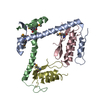
| ||||||||
|---|---|---|---|---|---|---|---|---|---|
| 1 |
| ||||||||
| Unit cell |
|
-Components
| #1: Protein | Mass: 11980.932 Da / Num. of mol.: 2 Source method: isolated from a genetically manipulated source Source: (gene. exp.) Shewanella oneidensis MR-1 (bacteria) / Strain: MR-1 / Gene: SO_1445 / Production host: #2: Protein | Mass: 11233.428 Da / Num. of mol.: 2 Source method: isolated from a genetically manipulated source Source: (gene. exp.) Shewanella oneidensis MR-1 (bacteria) / Strain: MR-1 / Gene: SO_1444 / Production host: Has ligand of interest | Y | Has protein modification | Y | |
|---|
-Experimental details
-Experiment
| Experiment | Method: X-RAY DIFFRACTION / Number of used crystals: 1 |
|---|
- Sample preparation
| Crystal | Density Matthews: 2.35 Å3/Da / Density % sol: 47.8 % |
|---|---|
| Crystal grow | Temperature: 293 K / Method: vapor diffusion, sitting drop / Details: 20% PEG3350 and 0.2 M tri-sodium citrate |
-Data collection
| Diffraction | Mean temperature: 100 K / Serial crystal experiment: N |
|---|---|
| Diffraction source | Source: SYNCHROTRON / Site: SSRF / Beamline: BL17U1 / Wavelength: 0.9788 Å |
| Detector | Type: DECTRIS PILATUS 6M / Detector: PIXEL / Date: May 26, 2019 |
| Radiation | Protocol: SINGLE WAVELENGTH / Monochromatic (M) / Laue (L): M / Scattering type: x-ray |
| Radiation wavelength | Wavelength: 0.9788 Å / Relative weight: 1 |
| Reflection | Resolution: 3.8→50 Å / Num. obs: 7720 / % possible obs: 100 % / Redundancy: 20 % / Biso Wilson estimate: 133 Å2 / CC1/2: 0.991 / Rmerge(I) obs: 0.195 / Rpim(I) all: 0.048 / Rrim(I) all: 0.205 / Net I/σ(I): 52 |
| Reflection shell | Resolution: 3.8→3.87 Å / Rmerge(I) obs: 0.796 / Mean I/σ(I) obs: 7.2 / Num. unique obs: 375 / CC1/2: 0.995 / Rpim(I) all: 0.155 / Rrim(I) all: 0.752 / % possible all: 100 |
- Processing
| Software |
| ||||||||||||||||||||||||||||||||||||||||||
|---|---|---|---|---|---|---|---|---|---|---|---|---|---|---|---|---|---|---|---|---|---|---|---|---|---|---|---|---|---|---|---|---|---|---|---|---|---|---|---|---|---|---|---|
| Refinement | Method to determine structure: SAD / Resolution: 3.804→33.93 Å / SU ML: 0.65 / Cross valid method: THROUGHOUT / σ(F): 1.41 / Phase error: 38.1 / Stereochemistry target values: ML
| ||||||||||||||||||||||||||||||||||||||||||
| Solvent computation | Shrinkage radii: 0.9 Å / VDW probe radii: 1.11 Å / Solvent model: FLAT BULK SOLVENT MODEL | ||||||||||||||||||||||||||||||||||||||||||
| Displacement parameters | Biso max: 310.63 Å2 / Biso mean: 138.358 Å2 / Biso min: 30 Å2 | ||||||||||||||||||||||||||||||||||||||||||
| Refinement step | Cycle: final / Resolution: 3.804→33.93 Å
| ||||||||||||||||||||||||||||||||||||||||||
| Refine LS restraints |
| ||||||||||||||||||||||||||||||||||||||||||
| LS refinement shell | Refine-ID: X-RAY DIFFRACTION / Rfactor Rfree error: 0
| ||||||||||||||||||||||||||||||||||||||||||
| Refinement TLS params. | Method: refined / Origin x: 88.5833 Å / Origin y: 53.4863 Å / Origin z: 18.3364 Å
| ||||||||||||||||||||||||||||||||||||||||||
| Refinement TLS group |
|
