 Movie
Movie Controller

[English] 日本語
 Yorodumi
- PDB-7esp: Structure and mutation analysis of the hexameric P4 from Pseudomo... -
+ Open data

- Basic information
| Entry | Database: PDB / ID: 7esp | ||||||
|---|---|---|---|---|---|---|---|
| Title | Structure and mutation analysis of the hexameric P4 from Pseudomonas aeruginosa phage phiYY | ||||||
 Components Components | Packaging NTPase | ||||||
 Keywords Keywords | HYDROLASE / Cystoviruses / phiYY P4 / NTPase / VIRAL PROTEIN | ||||||
| Function / homology | Packaging enzyme P4 / ATPase P4 of dsRNA bacteriophage phi-12 / viral genome packaging / P-loop containing nucleoside triphosphate hydrolase / metal ion binding / DI(HYDROXYETHYL)ETHER / D(-)-TARTARIC ACID / Packaging NTPase Function and homology information Function and homology information | ||||||
| Biological species |  Pseudomonas phage phiYY (virus) Pseudomonas phage phiYY (virus) | ||||||
| Method |  X-RAY DIFFRACTION / SYNCHROTRON / MOLECULAR REPLACEMENT / Resolution: 2.43 Å X-RAY DIFFRACTION / SYNCHROTRON / MOLECULAR REPLACEMENT / Resolution: 2.43 Å | ||||||
 Authors Authors | Zhang, C.Y. / Jin, T.C. | ||||||
 Citation Citation | Journal: Int.J.Biol.Macromol. / Year: 2022 Title: Structure and mutation analysis of the hexameric P4 from Pseudomonas aeruginosa phage phiYY. Authors: Zhang, C. / Li, Y. / Samad, A. / Zheng, P. / Ji, Z. / Chen, F. / Zhang, H. / Jin, T. | ||||||
| History |
|
- Structure visualization
| Structure viewer | Molecule: MolmilJmol/JSmol |
|---|
- Downloads & links
-Download
| PDBx/mmCIF format | 7esp.cif.gz | 400 KB | Display | PDBx/mmCIF format |
|---|---|---|---|---|
| PDB format | pdb7esp.ent.gz | 325.6 KB | Display | PDB format |
| PDBx/mmJSON format | 7esp.json.gz | Tree view | PDBx/mmJSON format | |
| Others |  Other downloads Other downloads |
-Validation report
| Summary document | 7esp_validation.pdf.gz | 2.9 MB | Display | wwPDB validaton report |
|---|---|---|---|---|
| Full document | 7esp_full_validation.pdf.gz | 2.9 MB | Display | |
| Data in XML | 7esp_validation.xml.gz | 77.6 KB | Display | |
| Data in CIF | 7esp_validation.cif.gz | 104.5 KB | Display | |
| Arichive directory | https://data.pdbj.org/pub/pdb/validation_reports/es/7espftp://data.pdbj.org/pub/pdb/validation_reports/es/7esp | HTTPS FTP |
-Related structure data
| Related structure data |  7esoC 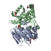 7esqC 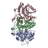 7esvC 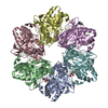 4blpS S: Starting model for refinement C: citing same article ( |
|---|---|
| Similar structure data |
-Links
 PDBj
PDBj
- Assembly
| Deposited unit | 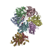
| ||||||||
|---|---|---|---|---|---|---|---|---|---|
| 1 | 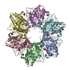
| ||||||||
| 2 | 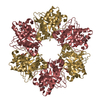
| ||||||||
| Unit cell |
|
-Components
| #1: Protein | Mass: 37338.531 Da / Num. of mol.: 8 Source method: isolated from a genetically manipulated source Source: (gene. exp.) Pseudomonas phage phiYY (virus) / Gene: phiYY_sL4 / Production host:  #2: Chemical | ChemComp-TAR /   Mass: 150.087 Da / Num. of mol.: 8 / Source method: obtained synthetically / Formula: C4H6O6 / Feature type: SUBJECT OF INVESTIGATION Mass: 150.087 Da / Num. of mol.: 8 / Source method: obtained synthetically / Formula: C4H6O6 / Feature type: SUBJECT OF INVESTIGATION#3: Chemical | ChemComp-PEG /   Mass: 106.120 Da / Num. of mol.: 4 / Source method: obtained synthetically / Formula: C4H10O3 Mass: 106.120 Da / Num. of mol.: 4 / Source method: obtained synthetically / Formula: C4H10O3#4: Water | ChemComp-HOH / |  Mass: 18.015 Da / Num. of mol.: 339 / Source method: isolated from a natural source / Formula: H2O Mass: 18.015 Da / Num. of mol.: 339 / Source method: isolated from a natural source / Formula: H2OHas ligand of interest | Y | |
|---|
-Experimental details
-Experiment
| Experiment | Method: X-RAY DIFFRACTION / Number of used crystals: 1 |
|---|
- Sample preparation
| Crystal | Density Matthews: 2.83 Å3/Da / Density % sol: 56.55 % |
|---|---|
| Crystal grow | Temperature: 291 K / Method: vapor diffusion, hanging drop / pH: 7.9 Details: 200 mM NH4 tartrate, 20% PEG2000 MME, phosphate pH 7.9 |
-Data collection
| Diffraction | Mean temperature: 100 K / Serial crystal experiment: N |
|---|---|
| Diffraction source | Source: SYNCHROTRON / Site: SSRF  / Beamline: BL17U1 / Wavelength: 0.979 Å / Beamline: BL17U1 / Wavelength: 0.979 Å |
| Detector | Type: DECTRIS EIGER X 16M / Detector: PIXEL / Date: Nov 23, 2017 |
| Radiation | Protocol: SINGLE WAVELENGTH / Monochromatic (M) / Laue (L): M / Scattering type: x-ray |
| Radiation wavelength | Wavelength: 0.979 Å / Relative weight: 1 |
| Reflection | Resolution: 2.43→40.97 Å / Num. obs: 1250846 / % possible obs: 97.8 % / Redundancy: 10.3 % / CC1/2: 0.998 / Net I/σ(I): 15.7 |
| Reflection shell | Resolution: 2.43→2.56 Å / Num. unique obs: 198187 / CC1/2: 0.834 |
- Processing
| Software |
| ||||||||||||||||||||||||||||||||||||||||||||||||||||||||||||||||||||||||||||||||||||||||||
|---|---|---|---|---|---|---|---|---|---|---|---|---|---|---|---|---|---|---|---|---|---|---|---|---|---|---|---|---|---|---|---|---|---|---|---|---|---|---|---|---|---|---|---|---|---|---|---|---|---|---|---|---|---|---|---|---|---|---|---|---|---|---|---|---|---|---|---|---|---|---|---|---|---|---|---|---|---|---|---|---|---|---|---|---|---|---|---|---|---|---|---|
| Refinement | Method to determine structure: MOLECULAR REPLACEMENT Starting model: 4blp Resolution: 2.43→40.97 Å / SU ML: 0.29 / Cross valid method: THROUGHOUT / σ(F): 2 / Phase error: 24.32 / Stereochemistry target values: ML
| ||||||||||||||||||||||||||||||||||||||||||||||||||||||||||||||||||||||||||||||||||||||||||
| Solvent computation | Shrinkage radii: 0.9 Å / VDW probe radii: 1.11 Å / Solvent model: FLAT BULK SOLVENT MODEL | ||||||||||||||||||||||||||||||||||||||||||||||||||||||||||||||||||||||||||||||||||||||||||
| Displacement parameters | Biso max: 113.52 Å2 / Biso mean: 61.5639 Å2 / Biso min: 34.79 Å2 | ||||||||||||||||||||||||||||||||||||||||||||||||||||||||||||||||||||||||||||||||||||||||||
| Refinement step | Cycle: final / Resolution: 2.43→40.97 Å
| ||||||||||||||||||||||||||||||||||||||||||||||||||||||||||||||||||||||||||||||||||||||||||
| LS refinement shell | Refine-ID: X-RAY DIFFRACTION / Rfactor Rfree error: 0
|
