 Movie
Movie Controller

[English] 日本語
 Yorodumi
- PDB-7eru: Crystal structure of L-histidine decarboxylase (C57S mutant) from... -
+ Open data

- Basic information
| Entry | Database: PDB / ID: 7eru | ||||||
|---|---|---|---|---|---|---|---|
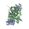
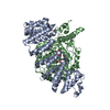
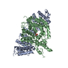
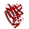
| Title | Crystal structure of L-histidine decarboxylase (C57S mutant) from Photobacterium phosphoreum | ||||||
 Components Components | Histidine decarboxylase | ||||||
 Keywords Keywords | LYASE / decarboxylase | ||||||
| Function / homology |  Function and homology information Function and homology informationhistidine decarboxylase / histidine decarboxylase activity / carboxylic acid metabolic process / pyridoxal phosphate binding Similarity search - Function | ||||||
| Biological species |  Photobacterium phosphoreum (bacteria) Photobacterium phosphoreum (bacteria) | ||||||
| Method |  X-RAY DIFFRACTION / SYNCHROTRON / MOLECULAR REPLACEMENT / Resolution: 2.85 Å X-RAY DIFFRACTION / SYNCHROTRON / MOLECULAR REPLACEMENT / Resolution: 2.85 Å | ||||||
 Authors Authors | Oda, Y. / Nakata, K. / Yamaguchi, H. / Kashiwagi, T. / Miyano, H. / Mizukoshi, T. | ||||||
 Citation Citation | Journal: J.Biochem. / Year: 2022 Title: Structural insights into the enhanced thermostability of cysteine substitution mutants of L-histidine decarboxylase from Photobacterium phosphoreum. Authors: Oda, Y. / Nakata, K. / Miyano, H. / Mizukoshi, T. / Yamaguchi, H. / Kashiwagi, T. | ||||||
| History |
|
- Structure visualization
| Structure viewer | Molecule: MolmilJmol/JSmol |
|---|
- Downloads & links
-Download
| PDBx/mmCIF format | 7eru.cif.gz | 89.1 KB | Display | PDBx/mmCIF format |
|---|---|---|---|---|
| PDB format | pdb7eru.ent.gz | 65.6 KB | Display | PDB format |
| PDBx/mmJSON format | 7eru.json.gz | Tree view | PDBx/mmJSON format | |
| Others |  Other downloads Other downloads |
-Validation report
| Summary document | 7eru_validation.pdf.gz | 431.2 KB | Display | wwPDB validaton report |
|---|---|---|---|---|
| Full document | 7eru_full_validation.pdf.gz | 437.7 KB | Display | |
| Data in XML | 7eru_validation.xml.gz | 15.5 KB | Display | |
| Data in CIF | 7eru_validation.cif.gz | 19.9 KB | Display | |
| Arichive directory | https://data.pdbj.org/pub/pdb/validation_reports/er/7eruftp://data.pdbj.org/pub/pdb/validation_reports/er/7eru | HTTPS FTP |
-Related structure data
| Related structure data | 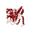 7ervC  3f9tS S: Starting model for refinement C: citing same article ( |
|---|---|
| Similar structure data |


-Links
 PDBj
PDBj- Assembly
| Deposited unit | 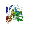
| ||||||||
|---|---|---|---|---|---|---|---|---|---|
| 1 | 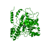
| ||||||||
| Unit cell |
|
-Components
| #1: Protein | Mass: 42190.547 Da / Num. of mol.: 1 / Mutation: C57S Source method: isolated from a genetically manipulated source Source: (gene. exp.) Photobacterium phosphoreum (bacteria) / Gene: hdc / Production host: |
|---|---|
| Has ligand of interest | Y |
-Experimental details
-Experiment
| Experiment | Method: X-RAY DIFFRACTION / Number of used crystals: 1 |
|---|
- Sample preparation
| Crystal | Density Matthews: 2.71 Å3/Da / Density % sol: 54.63 % |
|---|---|
| Crystal grow | Temperature: 293 K / Method: vapor diffusion, hanging drop / pH: 4.5 / Details: PEG1000, sodium acetate |
-Data collection
| Diffraction | Mean temperature: 100 K / Serial crystal experiment: N |
|---|---|
| Diffraction source | Source: SYNCHROTRON / Site: Photon Factory  / Beamline: BL-1A / Wavelength: 1.1 Å / Beamline: BL-1A / Wavelength: 1.1 Å |
| Detector | Type: DECTRIS EIGER X 4M / Detector: PIXEL / Date: Mar 3, 2017 |
| Radiation | Protocol: SINGLE WAVELENGTH / Monochromatic (M) / Laue (L): M / Scattering type: x-ray |
| Radiation wavelength | Wavelength: 1.1 Å / Relative weight: 1 |
| Reflection | Resolution: 2.85→100 Å / Num. obs: 11390 / % possible obs: 99.9 % / Redundancy: 18.7 % / Rmerge(I) obs: 0.103 / Net I/σ(I): 27.7 |
| Reflection shell | Resolution: 2.85→2.93 Å / Num. unique obs: 11390 / CC1/2: 0.949 |
- Processing
| Software |
| ||||||||||||||||||||||||||||||||||||||||||||||||||||||||||||
|---|---|---|---|---|---|---|---|---|---|---|---|---|---|---|---|---|---|---|---|---|---|---|---|---|---|---|---|---|---|---|---|---|---|---|---|---|---|---|---|---|---|---|---|---|---|---|---|---|---|---|---|---|---|---|---|---|---|---|---|---|---|
| Refinement | Method to determine structure: MOLECULAR REPLACEMENT Starting model: 3F9T Resolution: 2.85→45.13 Å / Cor.coef. Fo:Fc: 0.94 / Cor.coef. Fo:Fc free: 0.893 / SU B: 14.1 / SU ML: 0.276 / Cross valid method: THROUGHOUT / σ(F): 0 / ESU R Free: 0.377 / Stereochemistry target values: MAXIMUM LIKELIHOOD Details: HYDROGENS HAVE BEEN ADDED IN THE RIDING POSITIONS U VALUES : REFINED INDIVIDUALLY
| ||||||||||||||||||||||||||||||||||||||||||||||||||||||||||||
| Solvent computation | Ion probe radii: 0.8 Å / Shrinkage radii: 0.8 Å / VDW probe radii: 1.2 Å / Solvent model: MASK | ||||||||||||||||||||||||||||||||||||||||||||||||||||||||||||
| Displacement parameters | Biso max: 202.56 Å2 / Biso mean: 57.263 Å2 / Biso min: 23.99 Å2
| ||||||||||||||||||||||||||||||||||||||||||||||||||||||||||||
| Refinement step | Cycle: final / Resolution: 2.85→45.13 Å
| ||||||||||||||||||||||||||||||||||||||||||||||||||||||||||||
| Refine LS restraints |
| ||||||||||||||||||||||||||||||||||||||||||||||||||||||||||||
| LS refinement shell | Resolution: 2.85→2.924 Å / Rfactor Rfree error: 0 / Total num. of bins used: 20
|
