 Movie
Movie Controller


+ Open data

- Basic information
| Entry | Database: PDB / ID: 7eql | ||||||
|---|---|---|---|---|---|---|---|
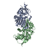
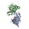
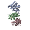
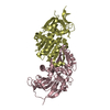
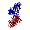
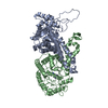
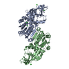
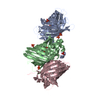
| Title | Crystal structure of (+)-pulegone reductase from Mentha piperita | ||||||
 Components Components | (+)-pulegone reductase | ||||||
 Keywords Keywords | PLANT PROTEIN / double bond reductase | ||||||
| Function / homology |  Function and homology information Function and homology information(+)-pulegone reductase / (+)-pulegone reductase (NADP+) activity / : / terpene metabolic process / terpenoid biosynthetic process / NADPH binding / cytoplasm Similarity search - Function | ||||||
| Biological species |  Mentha piperita (peppermint) Mentha piperita (peppermint) | ||||||
| Method |  X-RAY DIFFRACTION / SYNCHROTRON / MOLECULAR REPLACEMENT / Resolution: 2.72 Å X-RAY DIFFRACTION / SYNCHROTRON / MOLECULAR REPLACEMENT / Resolution: 2.72 Å | ||||||
 Authors Authors | Lin, W. | ||||||
 Citation Citation | Journal: Front Plant Sci / Year: 2021 Title: Functional Characterization and Structural Insights Into Stereoselectivity of Pulegone Reductase in Menthol Biosynthesis. Authors: Liu, C. / Gao, Q. / Shang, Z. / Liu, J. / Zhou, S. / Dang, J. / Liu, L. / Lange, I. / Srividya, N. / Lange, B.M. / Wu, Q. / Lin, W. | ||||||
| History |
|
- Structure visualization
| Structure viewer | Molecule: MolmilJmol/JSmol |
|---|
- Downloads & links
-Download
| PDBx/mmCIF format | 7eql.cif.gz | 79.3 KB | Display | PDBx/mmCIF format |
|---|---|---|---|---|
| PDB format | pdb7eql.ent.gz | 57.9 KB | Display | PDB format |
| PDBx/mmJSON format | 7eql.json.gz | Tree view | PDBx/mmJSON format | |
| Others |  Other downloads Other downloads |
-Validation report
| Summary document | 7eql_validation.pdf.gz | 429.3 KB | Display | wwPDB validaton report |
|---|---|---|---|---|
| Full document | 7eql_full_validation.pdf.gz | 440.6 KB | Display | |
| Data in XML | 7eql_validation.xml.gz | 14.2 KB | Display | |
| Data in CIF | 7eql_validation.cif.gz | 17.9 KB | Display | |
| Arichive directory | https://data.pdbj.org/pub/pdb/validation_reports/eq/7eqlftp://data.pdbj.org/pub/pdb/validation_reports/eq/7eql | HTTPS FTP |
-Related structure data
| Related structure data | 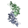 4hfjS S: Starting model for refinement |
|---|---|
| Similar structure data |


-Links
 PDBj
PDBj

- Assembly
| Deposited unit | 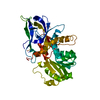
| ||||||||
|---|---|---|---|---|---|---|---|---|---|
| 1 | 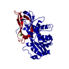
| ||||||||
| Unit cell |
|
-Components
| #1: Protein | (+)- Mass: 37952.719 Da / Num. of mol.: 1 Source method: isolated from a genetically manipulated source Source: (gene. exp.) Mentha piperita (peppermint)Production host:  References: UniProt: Q6WAU0, (+)-pulegone reductase |
|---|
-Experimental details
-Experiment
| Experiment | Method: X-RAY DIFFRACTION / Number of used crystals: 1 |
|---|
- Sample preparation
| Crystal | Density Matthews: 3.16 Å3/Da / Density % sol: 61.1 % Description: THE ENTRY CONTAINS FRIEDEL PAIRS IN I/F_PLUS/MINUS COLUMNS. |
|---|---|
| Crystal grow | Temperature: 293 K / Method: vapor diffusion, hanging drop / pH: 5.5 Details: 1M Ammonium sulfate , 0.1 M Bis-Tris (pH 5.5), 1% W/V PEG 3350 |
-Data collection
| Diffraction | Mean temperature: 100 K / Serial crystal experiment: N | ||||||||||||||||||||||||||||||
|---|---|---|---|---|---|---|---|---|---|---|---|---|---|---|---|---|---|---|---|---|---|---|---|---|---|---|---|---|---|---|---|
| Diffraction source | Source: SYNCHROTRON / Site: SSRF  / Beamline: BL17U / Wavelength: 0.97918 Å / Beamline: BL17U / Wavelength: 0.97918 Å | ||||||||||||||||||||||||||||||
| Detector | Type: DECTRIS PILATUS 6M / Detector: PIXEL / Date: Nov 19, 2019 | ||||||||||||||||||||||||||||||
| Radiation | Protocol: SINGLE WAVELENGTH / Monochromatic (M) / Laue (L): M / Scattering type: x-ray | ||||||||||||||||||||||||||||||
| Radiation wavelength | Wavelength: 0.97918 Å / Relative weight: 1 | ||||||||||||||||||||||||||||||
| Reflection | Resolution: 2.72→104.02 Å / Num. obs: 12444 / % possible obs: 96.3 % / Redundancy: 4 % / CC1/2: 0.999 / Rmerge(I) obs: 0.034 / Rpim(I) all: 0.019 / Rrim(I) all: 0.039 / Net I/σ(I): 19.6 / Num. measured all: 50025 | ||||||||||||||||||||||||||||||
| Reflection shell | Diffraction-ID: 1
|
- Processing
| Software |
| ||||||||||||||||||||||||||||||||||||
|---|---|---|---|---|---|---|---|---|---|---|---|---|---|---|---|---|---|---|---|---|---|---|---|---|---|---|---|---|---|---|---|---|---|---|---|---|---|
| Refinement | Method to determine structure: MOLECULAR REPLACEMENT Starting model: 4hfj Resolution: 2.72→104.019 Å / SU ML: 0.48 / Cross valid method: THROUGHOUT / σ(F): 1.38 / Phase error: 37.73 / Stereochemistry target values: ML
| ||||||||||||||||||||||||||||||||||||
| Solvent computation | Shrinkage radii: 0.9 Å / VDW probe radii: 1.11 Å / Solvent model: FLAT BULK SOLVENT MODEL | ||||||||||||||||||||||||||||||||||||
| Displacement parameters | Biso max: 170.61 Å2 / Biso mean: 112.1952 Å2 / Biso min: 20 Å2 | ||||||||||||||||||||||||||||||||||||
| Refinement step | Cycle: final / Resolution: 2.72→104.019 Å
| ||||||||||||||||||||||||||||||||||||
| LS refinement shell | Refine-ID: X-RAY DIFFRACTION / Rfactor Rfree error: 0
|
