 Movie
Movie Controller


+ Open data

- Basic information
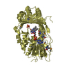
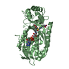
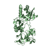
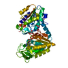
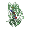
| Entry | Database: PDB / ID: 7epw | ||||||
|---|---|---|---|---|---|---|---|
| Title | Crystal structure of monooxygenase Tet(X4) with tigecycline | ||||||
 Components Components | Flavin-dependent monooxygenase | ||||||
 Keywords Keywords | OXIDOREDUCTASE / monooxygenase / Tet(X4) / tigecycline resistance | ||||||
| Function / homology |  Function and homology information Function and homology informationOxidoreductases; Acting on paired donors, with incorporation or reduction of molecular oxygen; With NADH or NADPH as one donor, and incorporation of one atom of oxygen into the other donor / FAD binding / monooxygenase activity / response to antibiotic / cytoplasm Similarity search - Function | ||||||
| Biological species |  | ||||||
| Method |  X-RAY DIFFRACTION / SYNCHROTRON / MOLECULAR REPLACEMENT / Resolution: 2.23 Å X-RAY DIFFRACTION / SYNCHROTRON / MOLECULAR REPLACEMENT / Resolution: 2.23 Å | ||||||
 Authors Authors | Cheng, Q. / Chen, S. | ||||||
 Citation Citation | Journal: Bmc Biol. / Year: 2021 Title: Structural and mechanistic basis of the high catalytic activity of monooxygenase Tet(X4) on tigecycline. Authors: Cheng, Q. / Cheung, Y. / Liu, C. / Xiao, Q. / Sun, B. / Zhou, J. / Chan, E.W.C. / Zhang, R. / Chen, S. | ||||||
| History |
|
- Structure visualization
| Structure viewer | Molecule: MolmilJmol/JSmol |
|---|
- Downloads & links
-Download
| PDBx/mmCIF format | 7epw.cif.gz | 95.2 KB | Display | PDBx/mmCIF format |
|---|---|---|---|---|
| PDB format | pdb7epw.ent.gz | 68.8 KB | Display | PDB format |
| PDBx/mmJSON format | 7epw.json.gz | Tree view | PDBx/mmJSON format | |
| Others |  Other downloads Other downloads |
-Validation report
| Arichive directory | https://data.pdbj.org/pub/pdb/validation_reports/ep/7epwftp://data.pdbj.org/pub/pdb/validation_reports/ep/7epw | HTTPS FTP |
|---|
-Related structure data
| Related structure data | 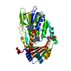 7epvSC S: Starting model for refinement C: citing same article ( |
|---|---|
| Similar structure data |


-Links
 PDBj
PDBj- Assembly
| Deposited unit | 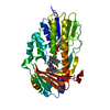
| ||||||||
|---|---|---|---|---|---|---|---|---|---|
| 1 |
| ||||||||
| Unit cell |
|
-Components
| #1: Protein | Mass: 43343.965 Da / Num. of mol.: 1 Source method: isolated from a genetically manipulated source Source: (gene. exp.) Production host: References: UniProt: A0A3T0V9Y5, Oxidoreductases; Acting on paired donors, with incorporation or reduction of molecular oxygen; With NADH or NADPH as one donor, and incorporation of one atom of ...References: UniProt: A0A3T0V9Y5, Oxidoreductases; Acting on paired donors, with incorporation or reduction of molecular oxygen; With NADH or NADPH as one donor, and incorporation of one atom of oxygen into the other donor |
|---|---|
| #2: Chemical | ChemComp-FDA /   Mass: 787.566 Da / Num. of mol.: 1 / Source method: obtained synthetically / Formula: C27H35N9O15P2 / Feature type: SUBJECT OF INVESTIGATION Mass: 787.566 Da / Num. of mol.: 1 / Source method: obtained synthetically / Formula: C27H35N9O15P2 / Feature type: SUBJECT OF INVESTIGATION |
| #3: Chemical | ChemComp-T1C / 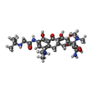  Mass: 587.665 Da / Num. of mol.: 1 / Source method: obtained synthetically / Formula: C29H41N5O8 / Feature type: SUBJECT OF INVESTIGATION / Comment: medication, antibiotic*YM Mass: 587.665 Da / Num. of mol.: 1 / Source method: obtained synthetically / Formula: C29H41N5O8 / Feature type: SUBJECT OF INVESTIGATION / Comment: medication, antibiotic*YM |
| #4: Water | ChemComp-HOH /  Mass: 18.015 Da / Num. of mol.: 77 / Source method: isolated from a natural source / Formula: H2O Mass: 18.015 Da / Num. of mol.: 77 / Source method: isolated from a natural source / Formula: H2O |
| Has ligand of interest | Y |
-Experimental details
-Experiment
| Experiment | Method: X-RAY DIFFRACTION / Number of used crystals: 1 |
|---|
- Sample preparation
| Crystal | Density Matthews: 2.67 Å3/Da / Density % sol: 53.92 % |
|---|---|
| Crystal grow | Temperature: 291 K / Method: vapor diffusion, sitting drop / pH: 5.6 Details: 30% PEG4000, 0.2 M Ammonium acetate, 0.1 M Sodium citrate tribasic dihydrate pH 5.6 PH range: 5.4-5.8 |
-Data collection
| Diffraction | Mean temperature: 100 K / Serial crystal experiment: N |
|---|---|
| Diffraction source | Source: SYNCHROTRON / Site: SSRF  / Beamline: BL17U1 / Wavelength: 0.979183 Å / Beamline: BL17U1 / Wavelength: 0.979183 Å |
| Detector | Type: DECTRIS EIGER X 16M / Detector: PIXEL / Date: Dec 30, 2020 |
| Radiation | Protocol: SINGLE WAVELENGTH / Monochromatic (M) / Laue (L): M / Scattering type: x-ray |
| Radiation wavelength | Wavelength: 0.979183 Å / Relative weight: 1 |
| Reflection | Resolution: 2.23→84.35 Å / Num. obs: 23785 / % possible obs: 99.6 % / Redundancy: 36.5 % / Rmerge(I) obs: 0.168 / Net I/σ(I): 22.5 |
| Reflection shell | Resolution: 2.23→2.29 Å / Rmerge(I) obs: 1.917 / Mean I/σ(I) obs: 2.3 / Num. unique obs: 1715 |
- Processing
| Software |
| ||||||||||||||||||||||||||||||||||||||||||||||||||||||||||||
|---|---|---|---|---|---|---|---|---|---|---|---|---|---|---|---|---|---|---|---|---|---|---|---|---|---|---|---|---|---|---|---|---|---|---|---|---|---|---|---|---|---|---|---|---|---|---|---|---|---|---|---|---|---|---|---|---|---|---|---|---|---|
| Refinement | Method to determine structure: MOLECULAR REPLACEMENT Starting model: 7EPV Resolution: 2.23→48.7 Å / Cor.coef. Fo:Fc: 0.963 / Cor.coef. Fo:Fc free: 0.931 / SU B: 6.131 / SU ML: 0.149 / Cross valid method: THROUGHOUT / σ(F): 0 / ESU R: 0.243 / ESU R Free: 0.209 / Stereochemistry target values: MAXIMUM LIKELIHOOD Details: HYDROGENS HAVE BEEN ADDED IN THE RIDING POSITIONS U VALUES : REFINED INDIVIDUALLY
| ||||||||||||||||||||||||||||||||||||||||||||||||||||||||||||
| Solvent computation | Ion probe radii: 0.8 Å / Shrinkage radii: 0.8 Å / VDW probe radii: 1.2 Å / Solvent model: MASK | ||||||||||||||||||||||||||||||||||||||||||||||||||||||||||||
| Displacement parameters | Biso max: 138.17 Å2 / Biso mean: 47.528 Å2 / Biso min: 25.84 Å2
| ||||||||||||||||||||||||||||||||||||||||||||||||||||||||||||
| Refinement step | Cycle: final / Resolution: 2.23→48.7 Å
| ||||||||||||||||||||||||||||||||||||||||||||||||||||||||||||
| Refine LS restraints |
| ||||||||||||||||||||||||||||||||||||||||||||||||||||||||||||
| LS refinement shell | Resolution: 2.23→2.288 Å / Rfactor Rfree error: 0 / Total num. of bins used: 20
|
