 Movie
Movie Controller

[English] 日本語
 Yorodumi
- PDB-7eoz: The structure of rice Defective Pollen Wall (DPW) in the complex ... -
+ Open data

- Basic information
| Entry | Database: PDB / ID: 7eoz | ||||||
|---|---|---|---|---|---|---|---|
| Title | The structure of rice Defective Pollen Wall (DPW) in the complex with its cofactor NADP | ||||||
 Components Components | Fatty acyl-CoA reductase | ||||||
 Keywords Keywords | OXIDOREDUCTASE / fatty acyl carrier protein reductase / UDP-glucose epimerase / lipid and sugar metabolisms / NADP+ / plant / LIPID BINDING PROTEIN | ||||||
| Function / homology |  Function and homology information Function and homology informationalcohol-forming fatty acyl-CoA reductase (NADPH) / alcohol-forming very long-chain fatty acyl-CoA reductase activity / alcohol-forming long-chain fatty acyl-CoA reductase activity / lipid metabolic process Similarity search - Function | ||||||
| Biological species |  | ||||||
| Method |  X-RAY DIFFRACTION / SYNCHROTRON / SAD / Resolution: 3.4 Å X-RAY DIFFRACTION / SYNCHROTRON / SAD / Resolution: 3.4 Å | ||||||
 Authors Authors | Yan, L.M. / Wang, W. / Li, G. / Wang, J. | ||||||
| Funding support |  China, 1items China, 1items
| ||||||
 Citation Citation | Journal: Plant Physiol Biochem. / Year: 2025 Title: Structural basis for the dual roles of DPW in lipid and UDP-sugar metabolism during rice anther development. Authors: Qu, S. / Wang, J. / Li, G. / Miao, C. / Yan, L. / Wang, W. | ||||||
| History |
|
- Structure visualization
| Structure viewer | Molecule: MolmilJmol/JSmol |
|---|
- Downloads & links
-Download
| PDBx/mmCIF format | 7eoz.cif.gz | 205.1 KB | Display | PDBx/mmCIF format |
|---|---|---|---|---|
| PDB format | pdb7eoz.ent.gz | 162 KB | Display | PDB format |
| PDBx/mmJSON format | 7eoz.json.gz | Tree view | PDBx/mmJSON format | |
| Others |  Other downloads Other downloads |
-Validation report
| Summary document | 7eoz_validation.pdf.gz | 1014.7 KB | Display | wwPDB validaton report |
|---|---|---|---|---|
| Full document | 7eoz_full_validation.pdf.gz | 1.1 MB | Display | |
| Data in XML | 7eoz_validation.xml.gz | 51.2 KB | Display | |
| Data in CIF | 7eoz_validation.cif.gz | 67.2 KB | Display | |
| Arichive directory | https://data.pdbj.org/pub/pdb/validation_reports/eo/7eozftp://data.pdbj.org/pub/pdb/validation_reports/eo/7eoz | HTTPS FTP |
-Related structure data
| Similar structure data |
|---|
-Links
 PDBj
PDBj- Assembly
| Deposited unit | 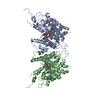
| ||||||||
|---|---|---|---|---|---|---|---|---|---|
| 1 | 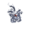
| ||||||||
| 2 | 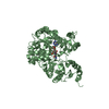
| ||||||||
| Unit cell |
|
-Components
| #1: Protein | Mass: 57963.586 Da / Num. of mol.: 2 Source method: isolated from a genetically manipulated source Source: (gene. exp.) Gene: OSJNBa0091P11.14, Os03g0167600 / Production host:  References: UniProt: Q0DUU1, alcohol-forming fatty acyl-CoA reductase (NADPH) #2: Chemical |   Mass: 743.405 Da / Num. of mol.: 2 / Source method: obtained synthetically / Formula: C21H28N7O17P3 / Feature type: SUBJECT OF INVESTIGATION Mass: 743.405 Da / Num. of mol.: 2 / Source method: obtained synthetically / Formula: C21H28N7O17P3 / Feature type: SUBJECT OF INVESTIGATION#3: Water | ChemComp-HOH / |  Mass: 18.015 Da / Num. of mol.: 85 / Source method: isolated from a natural source / Formula: H2O Mass: 18.015 Da / Num. of mol.: 85 / Source method: isolated from a natural source / Formula: H2OHas ligand of interest | Y | Has protein modification | N | |
|---|
-Experimental details
-Experiment
| Experiment | Method: X-RAY DIFFRACTION / Number of used crystals: 1 |
|---|
- Sample preparation
| Crystal grow | Temperature: 291 K / Method: evaporation Details: 2.8 M sodium formate, pH 7.0, 0.1 M magnesium chloride hexahydrate, 0.1 M HEPES, pH 7.5, 5% (w/v) PEG 3350 |
|---|
-Data collection
| Diffraction | Mean temperature: 93 K / Serial crystal experiment: N | |||||||||||||||||||||||||||||||||||||||||||||||||||||||||||||||||||||||||||||||||||||||||||||||||||||||||||||||||||||||||||||||||||||||||||||||||||
|---|---|---|---|---|---|---|---|---|---|---|---|---|---|---|---|---|---|---|---|---|---|---|---|---|---|---|---|---|---|---|---|---|---|---|---|---|---|---|---|---|---|---|---|---|---|---|---|---|---|---|---|---|---|---|---|---|---|---|---|---|---|---|---|---|---|---|---|---|---|---|---|---|---|---|---|---|---|---|---|---|---|---|---|---|---|---|---|---|---|---|---|---|---|---|---|---|---|---|---|---|---|---|---|---|---|---|---|---|---|---|---|---|---|---|---|---|---|---|---|---|---|---|---|---|---|---|---|---|---|---|---|---|---|---|---|---|---|---|---|---|---|---|---|---|---|---|---|---|
| Diffraction source | Source: SYNCHROTRON / Site: Photon Factory  / Beamline: BL-5A / Wavelength: 0.9798 Å / Beamline: BL-5A / Wavelength: 0.9798 Å | |||||||||||||||||||||||||||||||||||||||||||||||||||||||||||||||||||||||||||||||||||||||||||||||||||||||||||||||||||||||||||||||||||||||||||||||||||
| Detector | Type: ADSC QUANTUM 270 / Detector: CCD / Date: Mar 17, 2012 | |||||||||||||||||||||||||||||||||||||||||||||||||||||||||||||||||||||||||||||||||||||||||||||||||||||||||||||||||||||||||||||||||||||||||||||||||||
| Radiation | Protocol: SINGLE WAVELENGTH / Monochromatic (M) / Laue (L): M / Scattering type: x-ray | |||||||||||||||||||||||||||||||||||||||||||||||||||||||||||||||||||||||||||||||||||||||||||||||||||||||||||||||||||||||||||||||||||||||||||||||||||
| Radiation wavelength | Wavelength: 0.9798 Å / Relative weight: 1 | |||||||||||||||||||||||||||||||||||||||||||||||||||||||||||||||||||||||||||||||||||||||||||||||||||||||||||||||||||||||||||||||||||||||||||||||||||
| Reflection | Resolution: 3.3→50 Å / Num. obs: 48784 / % possible obs: 99.8 % / Redundancy: 11.1 % / Rmerge(I) obs: 0.136 / Χ2: 5.755 / Net I/σ(I): 16 / Num. measured all: 540754 | |||||||||||||||||||||||||||||||||||||||||||||||||||||||||||||||||||||||||||||||||||||||||||||||||||||||||||||||||||||||||||||||||||||||||||||||||||
| Reflection shell |
|
- Processing
| Software |
| ||||||||||||||||||||||||||||||||||||||||||||||||||||||||||||
|---|---|---|---|---|---|---|---|---|---|---|---|---|---|---|---|---|---|---|---|---|---|---|---|---|---|---|---|---|---|---|---|---|---|---|---|---|---|---|---|---|---|---|---|---|---|---|---|---|---|---|---|---|---|---|---|---|---|---|---|---|---|
| Refinement | Method to determine structure: SAD / Resolution: 3.4→48.45 Å / Cor.coef. Fo:Fc: 0.935 / Cor.coef. Fo:Fc free: 0.907 / SU B: 15.94 / SU ML: 0.243 / Cross valid method: THROUGHOUT / σ(F): 0 / ESU R: 0.559 / ESU R Free: 0.359 / Stereochemistry target values: MAXIMUM LIKELIHOOD Details: HYDROGENS HAVE BEEN ADDED IN THE RIDING POSITIONS U VALUES : REFINED INDIVIDUALLY
| ||||||||||||||||||||||||||||||||||||||||||||||||||||||||||||
| Solvent computation | Ion probe radii: 0.8 Å / Shrinkage radii: 0.8 Å / VDW probe radii: 1.2 Å / Solvent model: MASK | ||||||||||||||||||||||||||||||||||||||||||||||||||||||||||||
| Displacement parameters | Biso max: 264.51 Å2 / Biso mean: 94.95 Å2 / Biso min: 30 Å2
| ||||||||||||||||||||||||||||||||||||||||||||||||||||||||||||
| Refinement step | Cycle: final / Resolution: 3.4→48.45 Å
| ||||||||||||||||||||||||||||||||||||||||||||||||||||||||||||
| Refine LS restraints |
| ||||||||||||||||||||||||||||||||||||||||||||||||||||||||||||
| LS refinement shell | Resolution: 3.4→3.488 Å / Rfactor Rfree error: 0 / Total num. of bins used: 20
|
